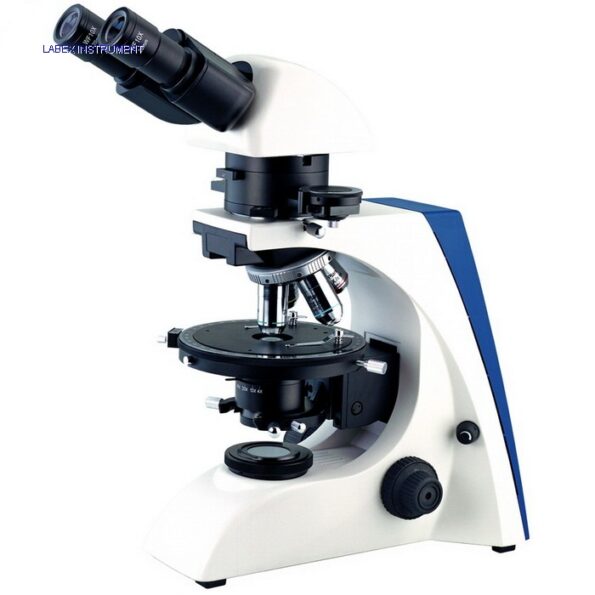
LABEX EUM-5000P Polarization Microscope
AED 10,185.00
Polarizing microscope LABEX EUM-5000P offers high-definition image quality in polarized light, using contrast-enhancing technique. This microscope comes with quintuple click stop revolving mechanism with multiple ball bearing. This polarizing microscope has Siedentop type 30° inclined binocular head. It can be equipped with filters e.g. blue, ground glass for more even and diffused light.
Available on backorder
Description
Used to study polarized light effect on a specimen in the field of science, forensic analysis, geology, medicine, chemistry, biology & metallurgy.
Polarizing Microscope
Polarizing microscopes are designed to image samples which are visible primarily due to optical anisotropy, or measurable differences in refractivity. Over 90% of solid samples exhibit optical properties that vary with the orientation of incident light and are therefore amenable to imaging. The technique offers significant advantages for samples that exhibit poor phase contrast by other means so as light or fluorescence. It can also reveal details concerning the structure and composition of materials.
Components including the polarizer and the analyzer are crucial to performance and quality should be considered accordingly. The light source and the lens objectives require consideration, as do the digital camera system and epi-illuminator components, if applicable.
| Viewing head | Siedentopf type binocular head, 30° inclined, eyepiece rotation 360° Interpupillary adjustable distance: 50 to 75 mm |
| Eyepiece | WF10X/20 mm, WF10X/20 mm (reticle 0.1 mm) |
| Objectives | Non-stress infinite plan objective: 4x, 10x, 20x, 40x(s) |
| Nosepiece | Quintuple click stop revolving mechanism with multiple ball bearing |
| Stage | Rotable 360°, center adjustable, division 1°, vernier division 6’ |
| Condenser | Swing type N.A. 1.25 with iris diaphragm and filter |
| Focusing | Coaxial coarse & fine adjustment, focusing range 30 mm, focusing interval 0.002 mm |
| light source | 6V/20W Halogen lamp, brightness adjustable |



0.0 Average Rating Rated (0 Reviews)