Key Features
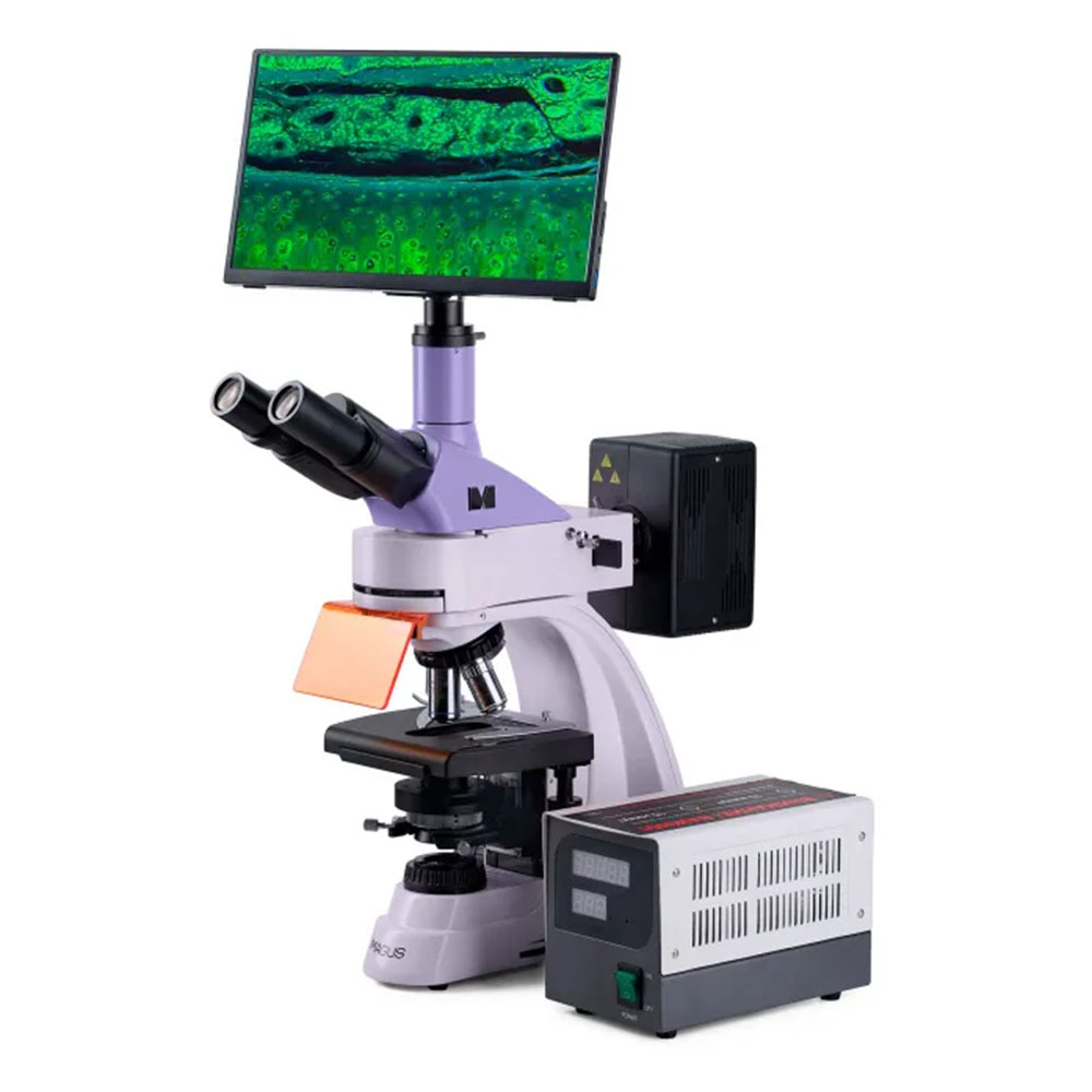
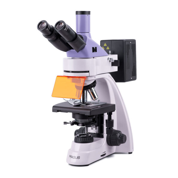
High-Power Trinocular Microscope: 40x - 1000x Magnification
- Detailed Observations: Explore microscopic worlds with exceptional 40x to 1000x magnification.
- Versatile Imaging: Capture stunning images and videos with the included camera and monitor. (Optional: Add a note about camera compatibility if applicable)
- Superior Optics: Trinocular head design allows for comfortable viewing and simultaneous camera attachment.
- Crystal-Clear Clarity: Plan achromatic and plan fluorite objectives deliver sharp, high-resolution images.
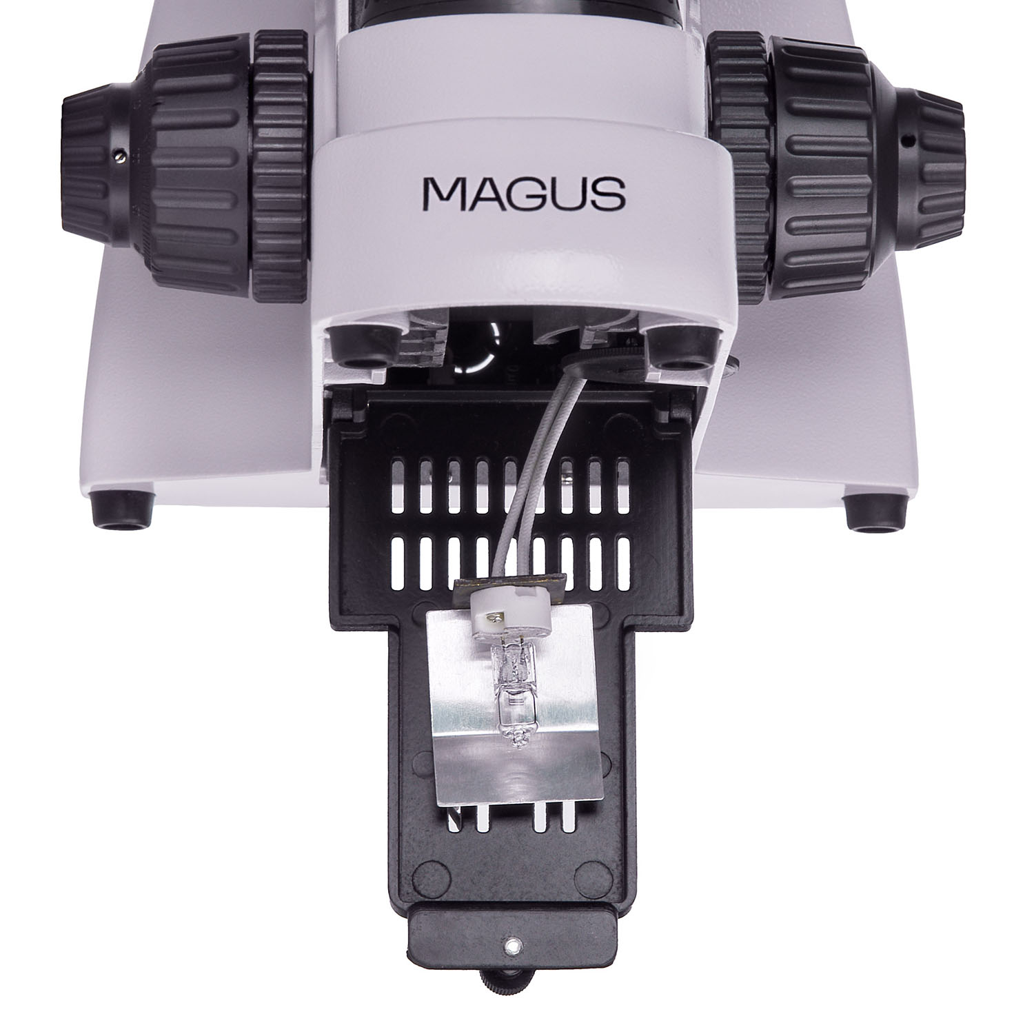
- Dual Illumination: Choose between reflected (100W mercury lamp) and transmitted (30W halogen lamp) light sources for optimal sample visualization under various conditions.
- Köhler Illumination: Ensures even and consistent lighting for accurate analysis
Description
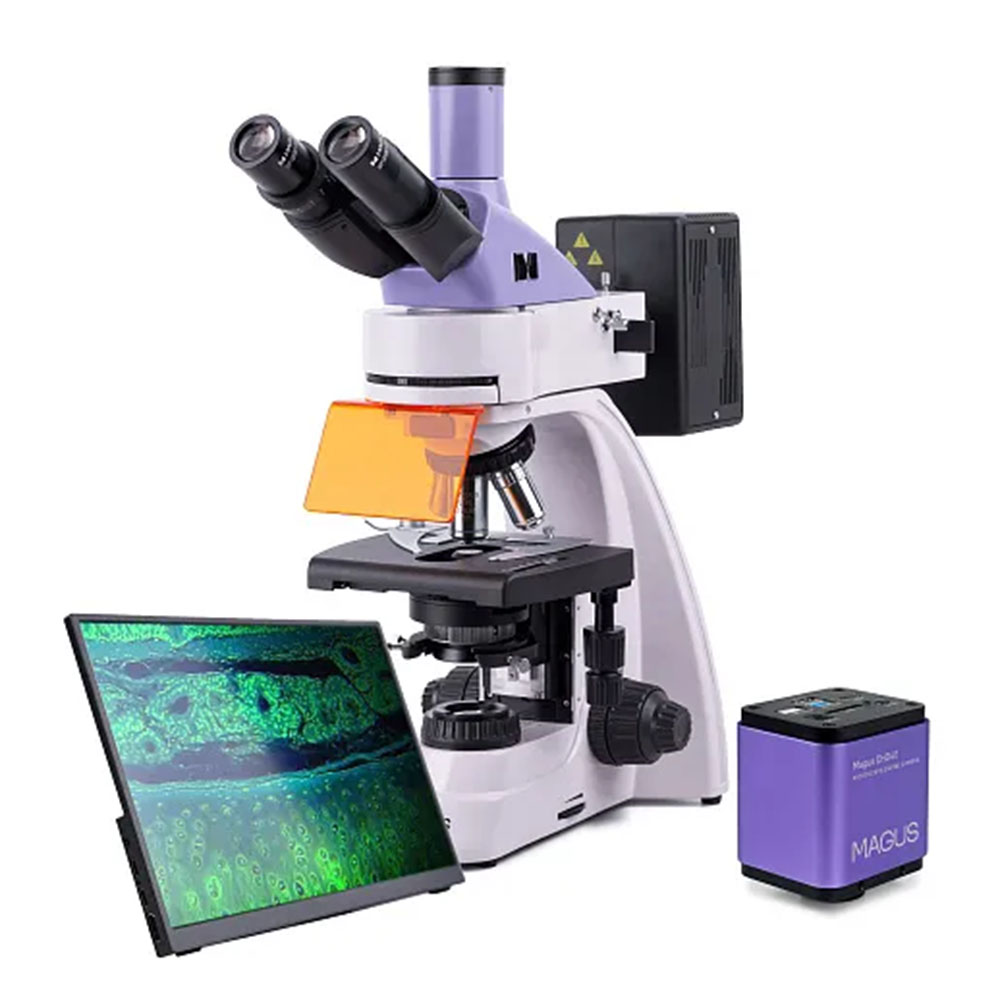
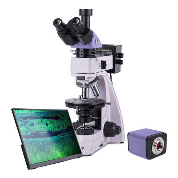
Unveiling the Microscopic World: MAGUS Lum 400 Fluorescence Microscope
See Beyond the Visible: Powerful Magnification for Diverse Research Needs
The MAGUS Lum 400 fluorescence microscope empowers researchers and diagnosticians to unlock the secrets of the microscopic world. This versatile instrument offers exceptional magnification capabilities (40x - 1000x) for detailed observations across various applications.
Fluorescence & Brightfield Techniques in One:
- Fluorescence microscopy (reflected light): Ideal for visualizing fluorescently labeled specimens, perfect for DNA analysis, pathogen identification, and more.
- Brightfield microscopy (transmitted light): Provides clear observations of a wider range of samples, suitable for routine diagnostic procedures and general cell studies.
Expandable Functionality (Optional Accessories):
- Phase contrast microscopy: Enhances contrast for transparent samples, revealing fine cellular details.
- Darkfield microscopy: Enables observation of highly transparent or unstained specimens, ideal for studying bacteria and microorganisms.
- Polarization microscopy: Analyzes birefringent materials, a valuable tool for crystal and mineral research.
Capture Stunning Visual Documentation (Optional Camera):
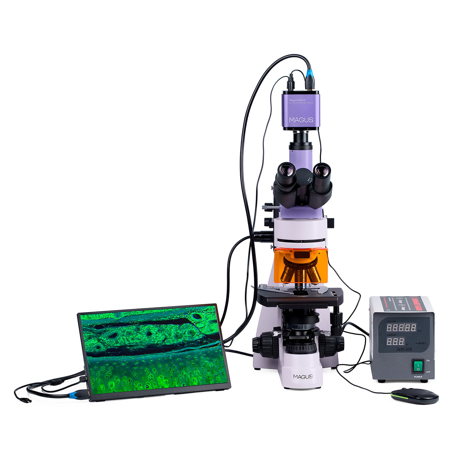
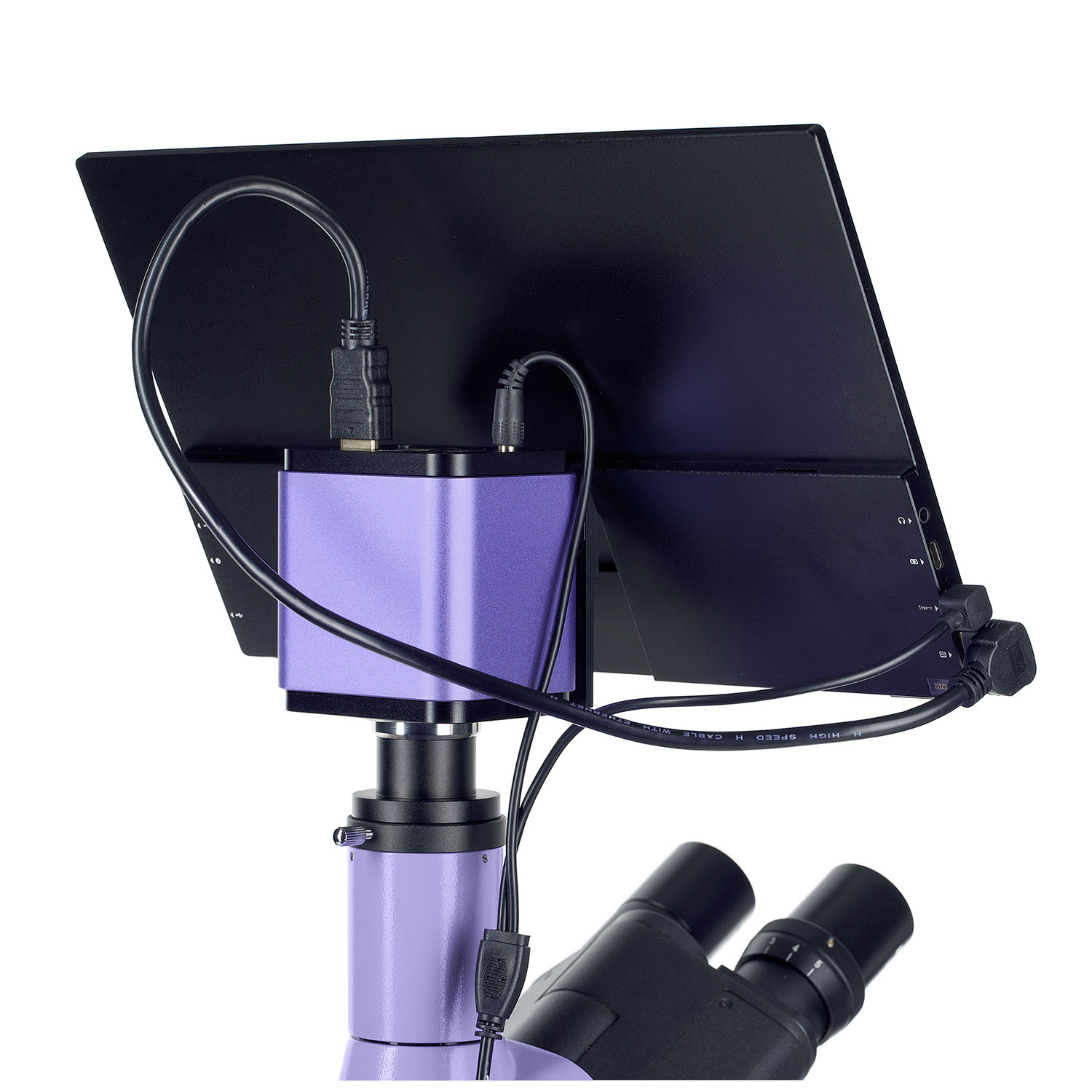
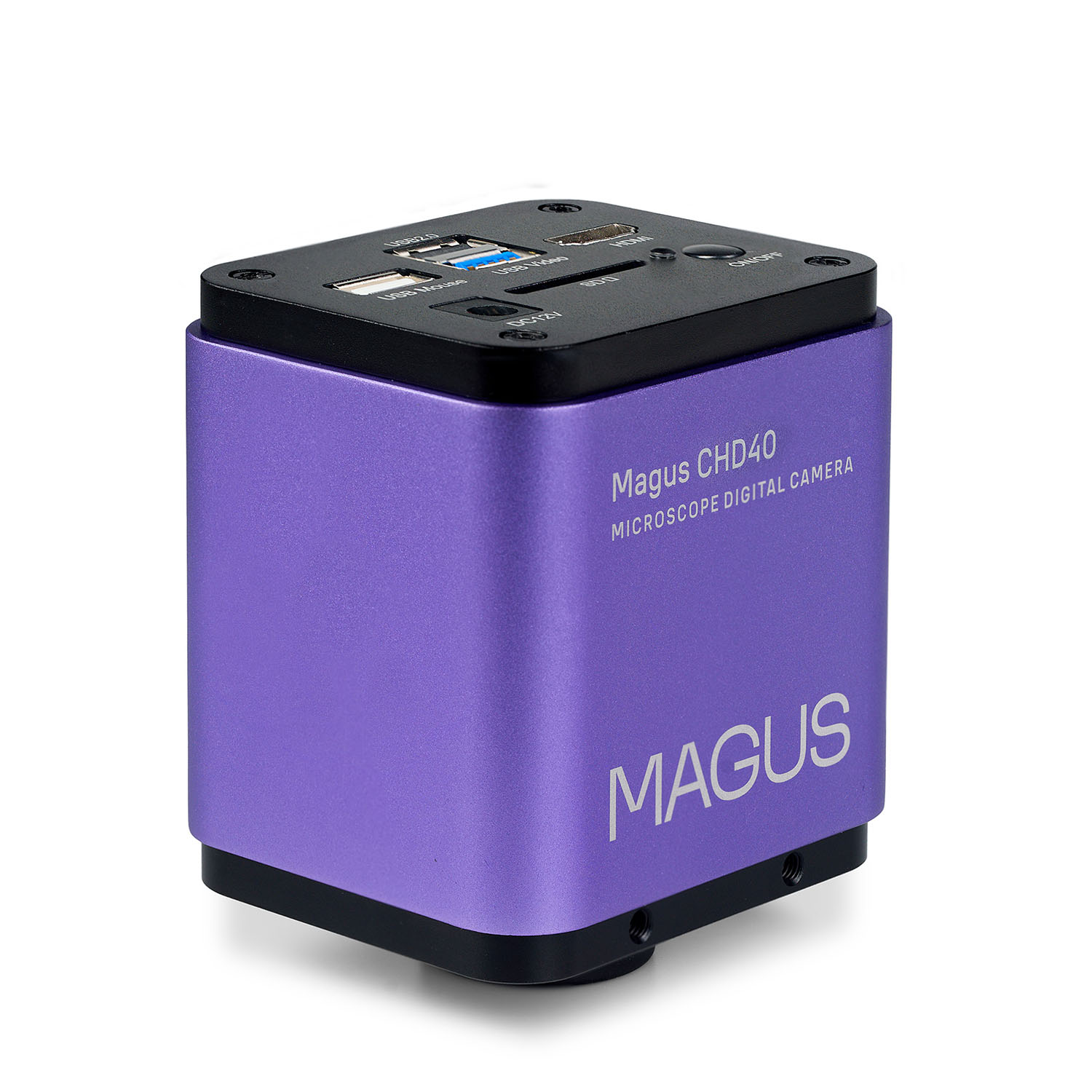
- MAGUS CHD40 Digital Camera: Seamlessly integrates with the microscope for high-resolution image and video capture (up to 4K resolution).
- Exceptional Low-Light Performance: Delivers crisp, clear images even in challenging lighting conditions.
Advanced Optical Performance:
- Infinity plan achromatic objectives: Deliver superior image clarity and resolution.
- Dual Illumination: Powerful 100W mercury lamp for fluorescence and 30W halogen lamp for brightfield microscopy ensures optimal sample visualization.
- Köhler Illumination: Provides even and consistent lighting for accurate analysis and minimizes image artifacts.
Ergonomic Design for Comfort:
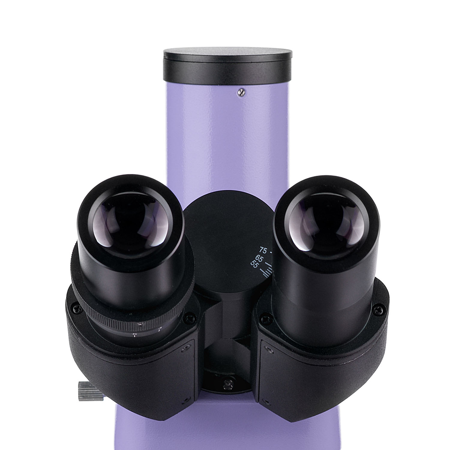
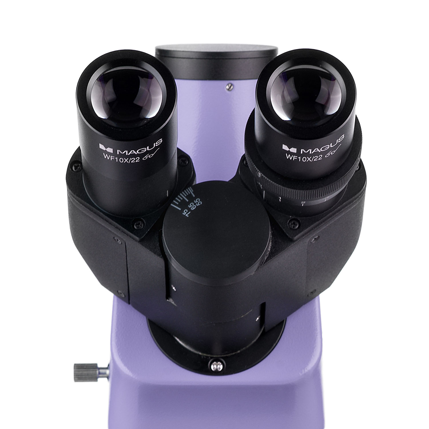
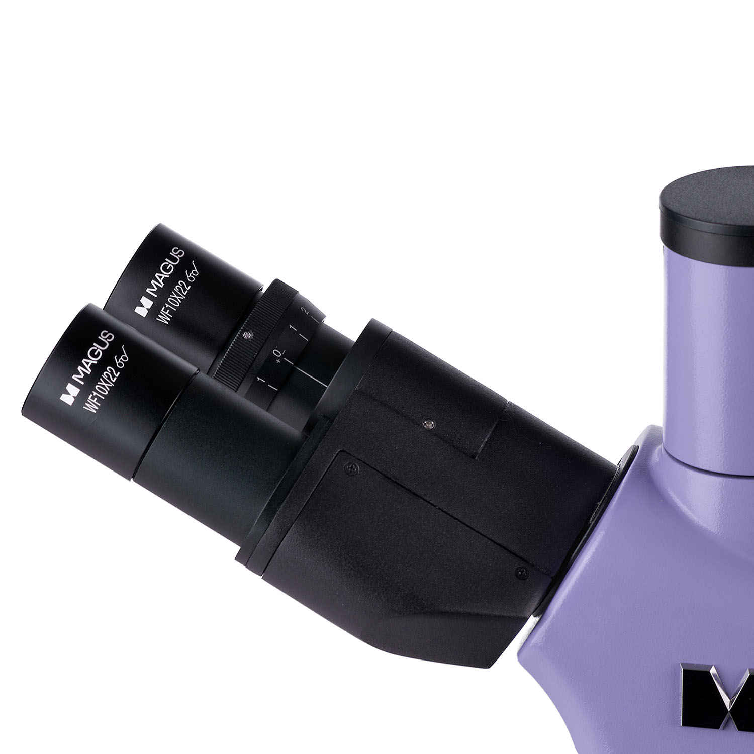
- 30° Inclined Binocular Head: Reduces neck strain during extended observation sessions.
- 360° Rotatable Eyepieces: Allow for personalized adjustments for optimal comfort.
- Ergonomically Designed Stage: Simplifies sample manipulation and streamlines workflow.
Complete System, Ready to Explore:
The MAGUS Lum 400 arrives as a complete system, including everything you need to begin your microscopic investigations. A variety of optional accessories further extend your research capabilities.
Unleash the Power of Microscopy: Explore the unseen world with the MAGUS Lum 400 fluorescence microscope.
Microscope Highlights:
- Unveiling a Hidden World: Multiple Microscopy Techniques: The MAGUS Lum 400 empowers you with a versatile range of microscopy techniques. Explore the world of fluorescently labeled specimens using fluorescence microscopy (reflected light), ideal for applications like DNA analysis and pathogen identification. Brightfield microscopy (transmitted light) provides detailed observations of a wider range of samples, making it suitable for routine diagnostic procedures and general cell studies. Don't be limited by built-in functionalities - the microscope can be expanded to encompass even more with optional accessories (sold separately). Incorporate phase contrast microscopy to enhance the contrast of transparent samples, revealing fine cellular details often invisible under brightfield illumination. Delve into the world of darkfield microscopy for observing highly transparent or unstained specimens, a valuable tool for studying bacteria and other microorganisms. Explore the properties of birefringent materials with polarization microscopy, a technique useful for analyzing crystals and minerals.
- Superior Optics for Exceptional Image Clarity: Infinity plan achromatic objectives are the cornerstone of the MAGUS Lum 400's optical system, renowned for their crispness and high resolution. This translates to sharp, detailed images that allow you to confidently analyze even the most minute cellular structures. Dual illumination ensures optimal sample visualization under various conditions. The powerful 100W mercury lamp provides the excitation light necessary for fluorescence microscopy, while the 30W halogen lamp offers bright illumination for brightfield observation. Köhler illumination, a cornerstone of microscopy, is achievable with this microscope. This method delivers even and consistent lighting across the sample, minimizing image artifacts and ensuring accurate analysis.
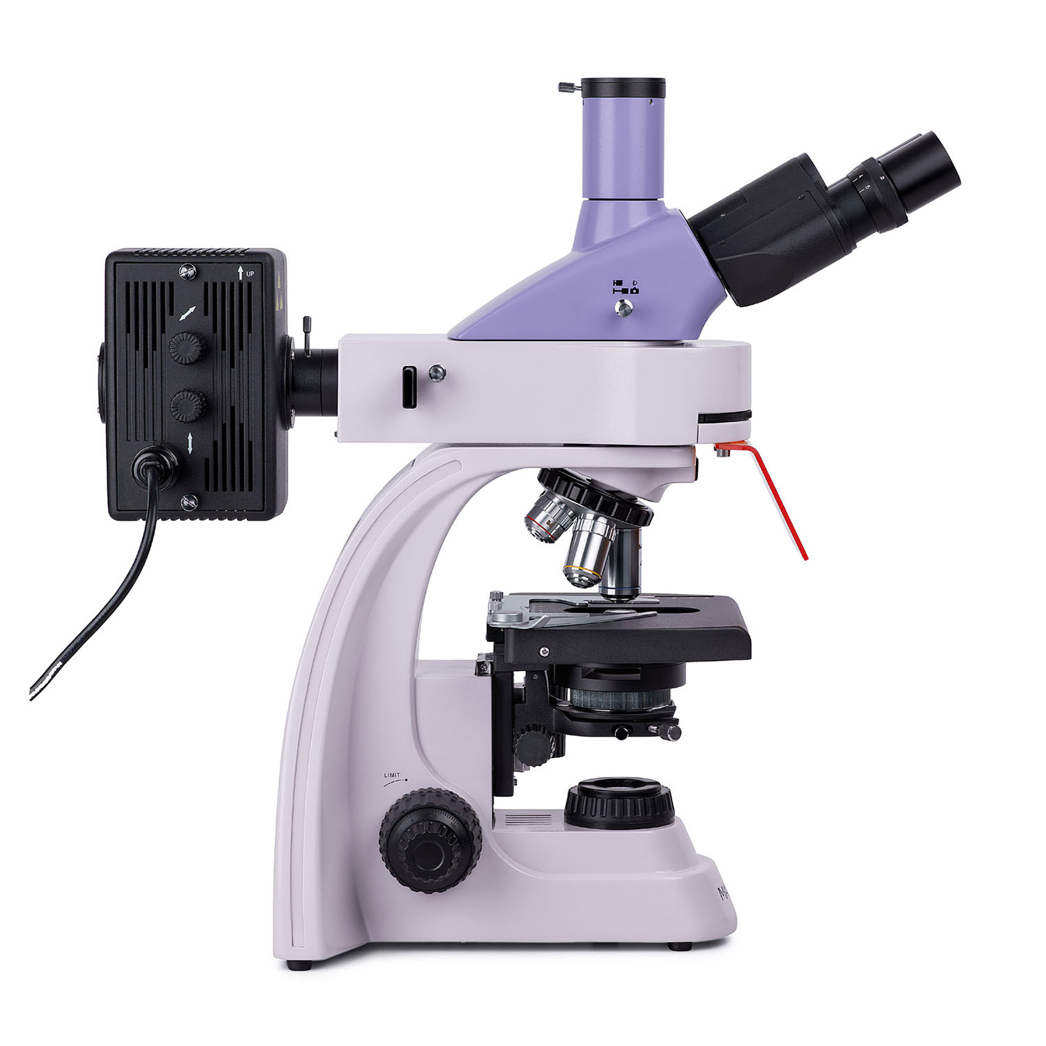
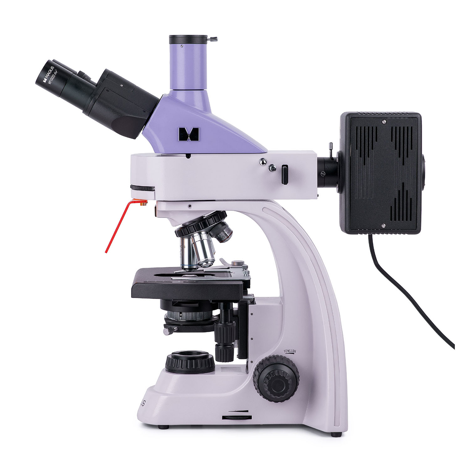
- Designed for Comfort and Efficiency: Extended observation sessions are a breeze with the MAGUS Lum 400's ergonomic design. The 30° inclined trinocular head minimizes neck strain, allowing you to comfortably focus on your research for longer durations. Rotatable eyepieces provide personalized adjustments for optimal viewing, ensuring a comfortable experience for users of various heights. The ergonomically designed stage simplifies sample manipulation and streamlines your workflow, allowing you to focus on making critical observations and discoveries.
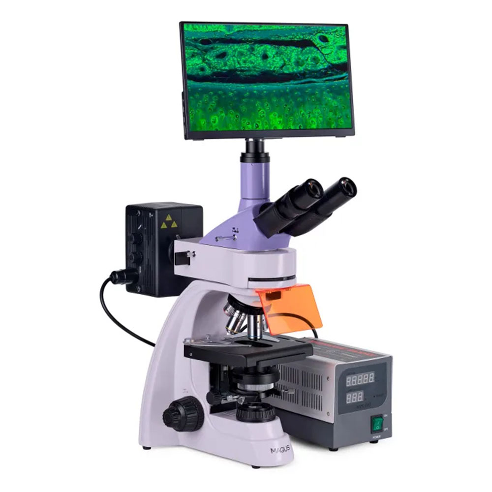
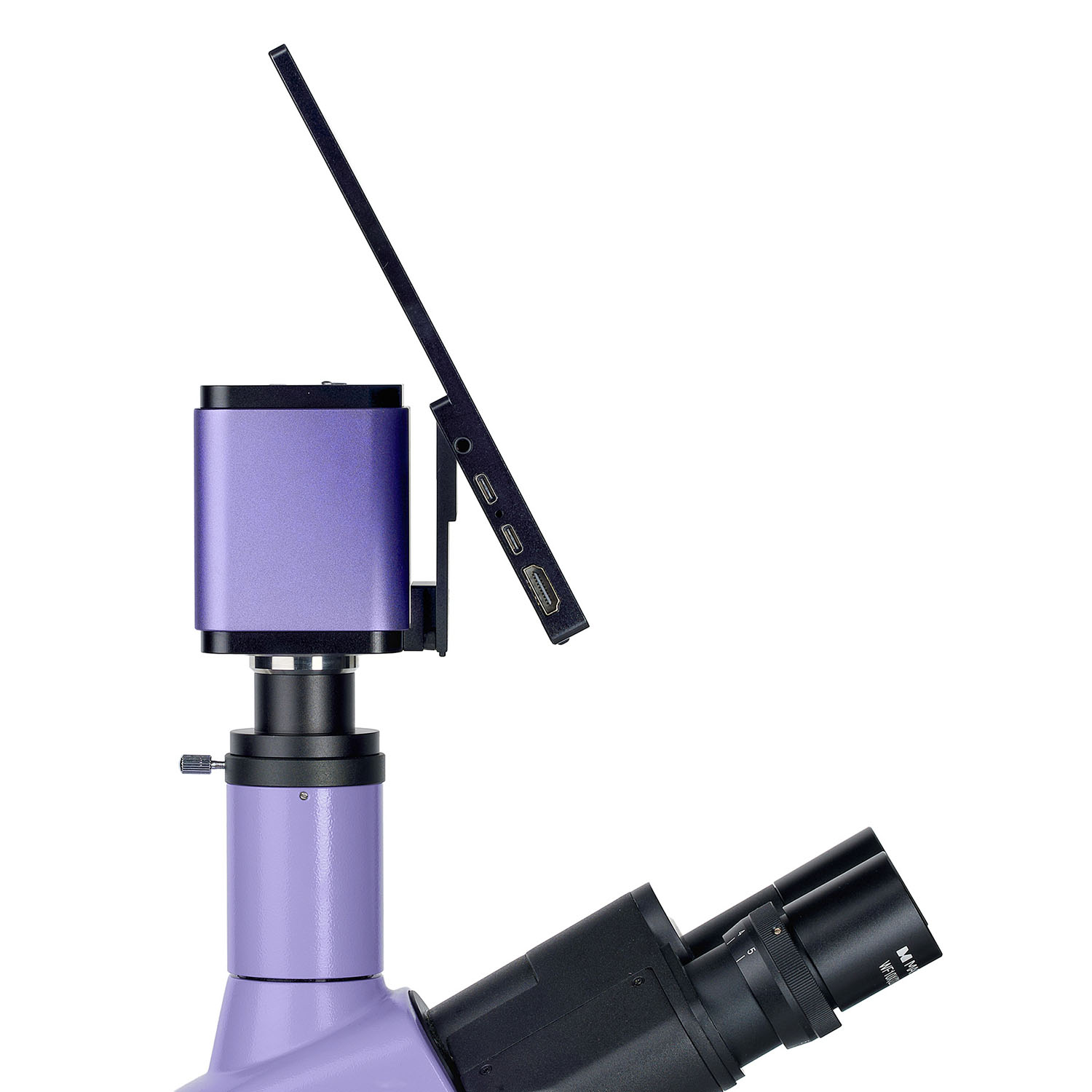
- Seamless Digital Integration for Enhanced Documentation (Optional Camera): Elevate your research and share discoveries with ease by incorporating the optional MAGUS CHD40 Digital Camera. This versatile camera seamlessly integrates with the trinocular head, allowing for simultaneous viewing and high-resolution image and video capture (up to 4K resolution). No matter the lighting conditions, the camera boasts exceptional low-light performance thanks to the SONY Exmor/Starvis sensor. This ensures crisp and clear image capture, even in challenging laboratory environments. The included software offers functionalities for photo and video editing, external display functions, and linear and angular measurements, empowering you to create comprehensive reports and presentations.
Camera Highlights:
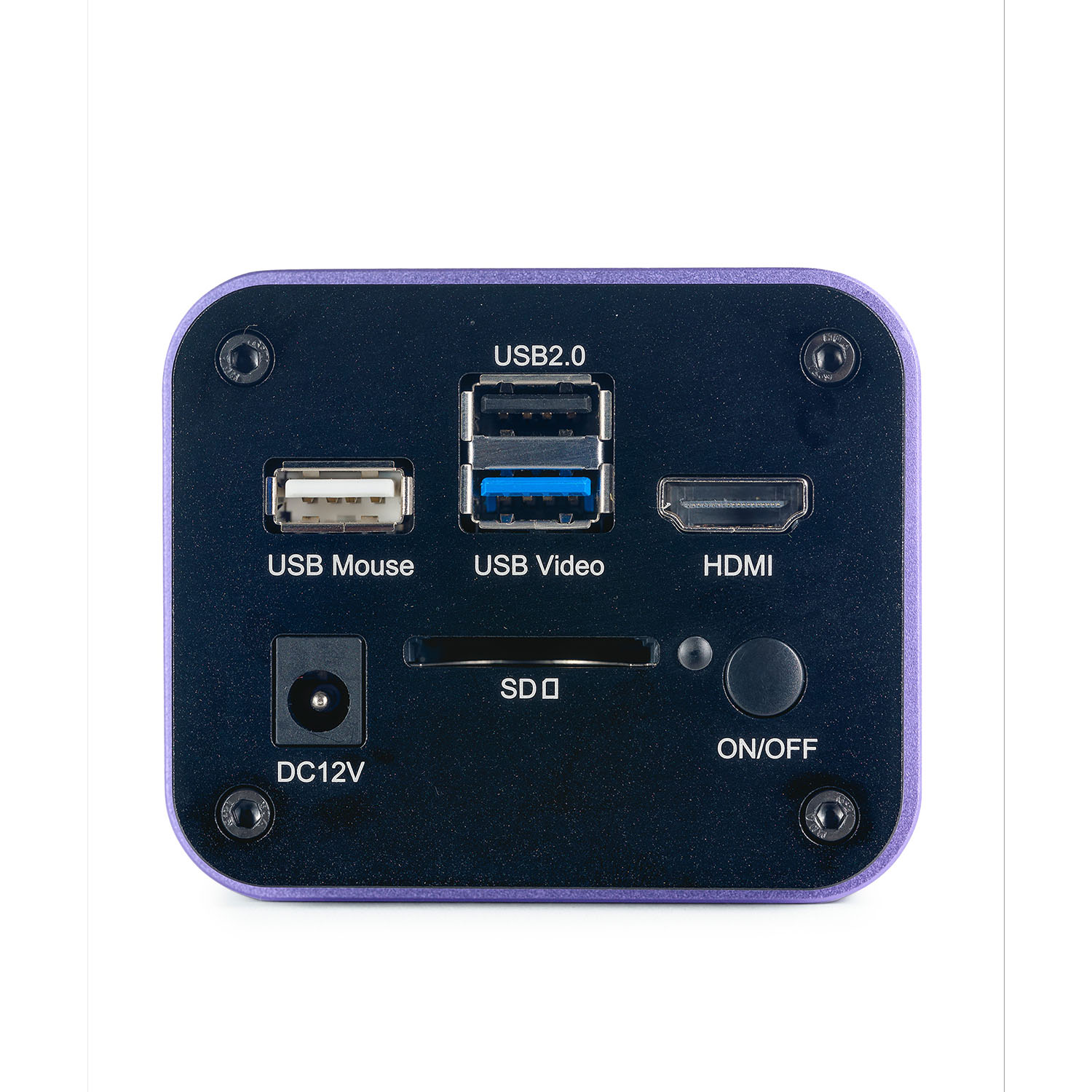
- Versatile Connectivity: The MAGUS CHD40 Digital Camera (optional) offers exceptional flexibility in how you connect and utilize it. Operate the camera independently for on-the-go image capture, or connect it to your PC via Wi-Fi or USB3.0 for seamless integration with your workflow. For presentations or sharing your findings on a larger screen, the HDMI interface allows for direct connection to TVs, monitors, or projectors.
- Stunning Image Quality with Auto-Switching Resolution: Capture high-quality images and videos that leave a lasting impression. The camera boasts auto-switching resolution between 4K and Full HD depending on the monitor you connect to, ensuring optimal image quality for the intended display. The SONY Exmor/Starvis sensor plays a crucial role once again, delivering exceptional low-light performance. This translates to crisp and clear images, even in dimly lit laboratory settings.
- Smooth Video Recording for Detailed Analysis: Record smooth, jerk-free videos at 30fps, ideal for capturing the dynamics of biological processes or observing the movement of microorganisms within a sample. This allows for detailed analysis of even the most subtle changes, providing valuable insights into your research endeavors.
Comprehensive Microscope Kit:
The MAGUS Lum 400 arrives as a complete system, ready to use right out of the box. It includes everything you need to begin your microscopic investigations, including the microscope itself, a trinocular head, eyepieces, objectives, illumination system, and stage. A variety of optional accessories further extend your research capabilities, allowing you to tailor the microscope to your specific needs. These optional accessories include the aforementioned digital camera, phase contrast device, polarization device, darkfield condenser, additional eyepieces and objectives, and a calibration slide for precise measurements.
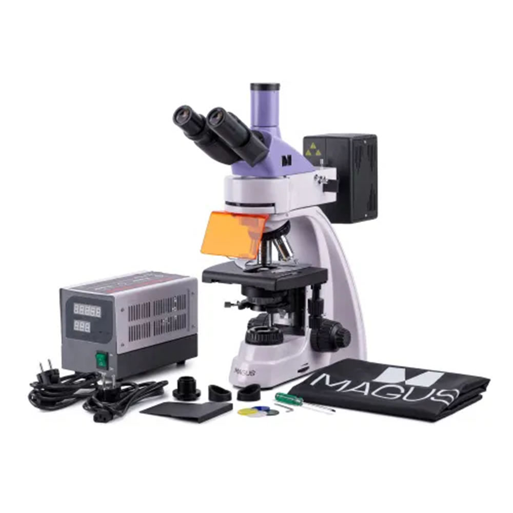
The kit includes:
- MAGUS CHD40 Digital Camera (digital camera, HDMI cable (1.5m), USB3.0 cable (1.5m), USB mouse, 32GB SD memory card, USB Wi-Fi adapter (2pcs.), AC power adapter 12V/1A (Euro), installation CD with drivers and software, user manual and warranty card)
- MAGUS MCD40 LCD Monitor
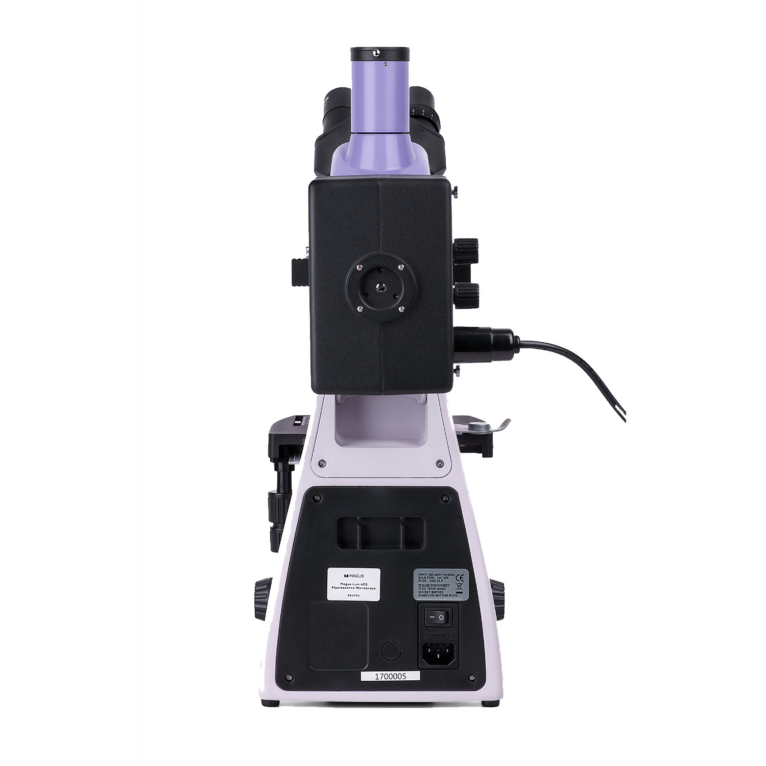
- Base with a power input, transmitted light source and condenser, focusing mechanism, stage, and revolving nosepiece
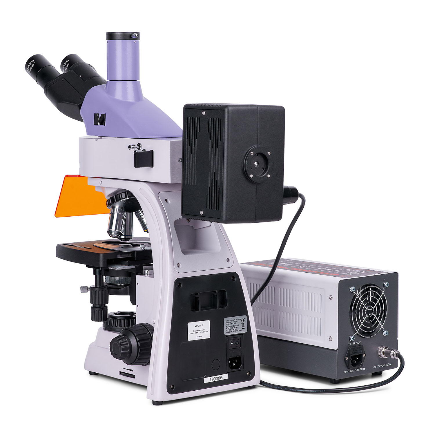
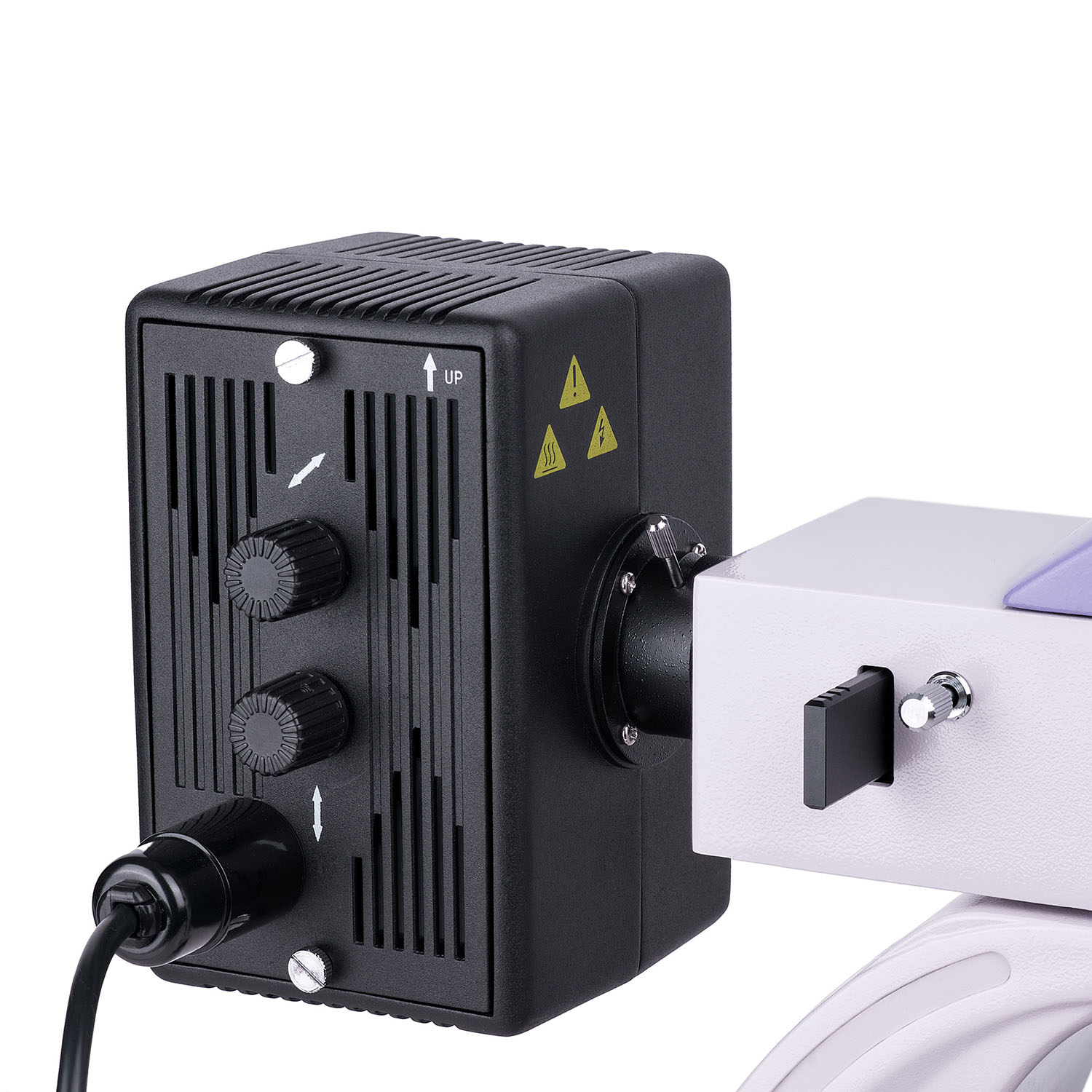
- Reflected light illuminator
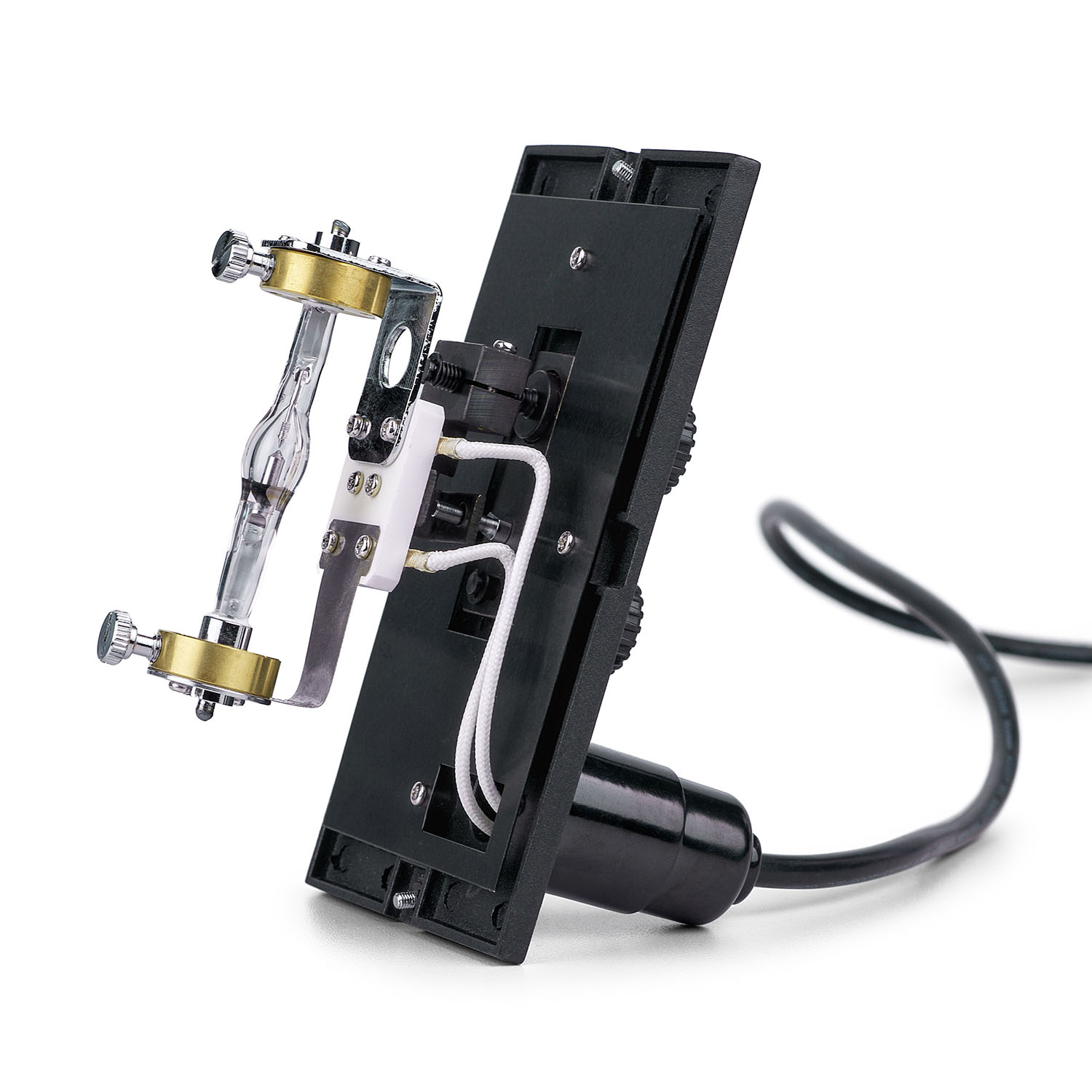
- Mercury lamphouse
- Trinocular head
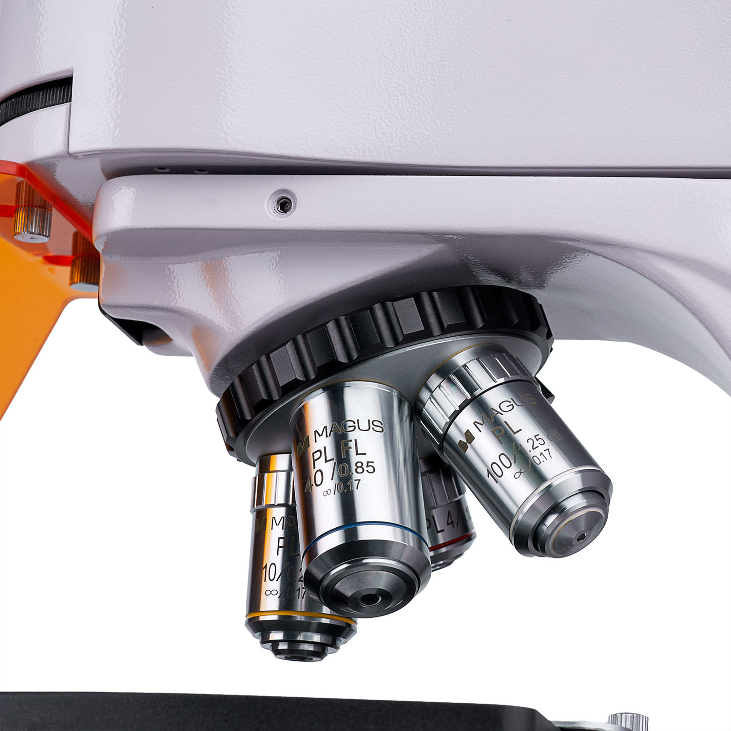
- Infinity plan achromatic objective: PL 4x/0.10 WD 19.8mm
- Infinity plan achromatic objective: PL 10x/0.25 WD 5.0mm
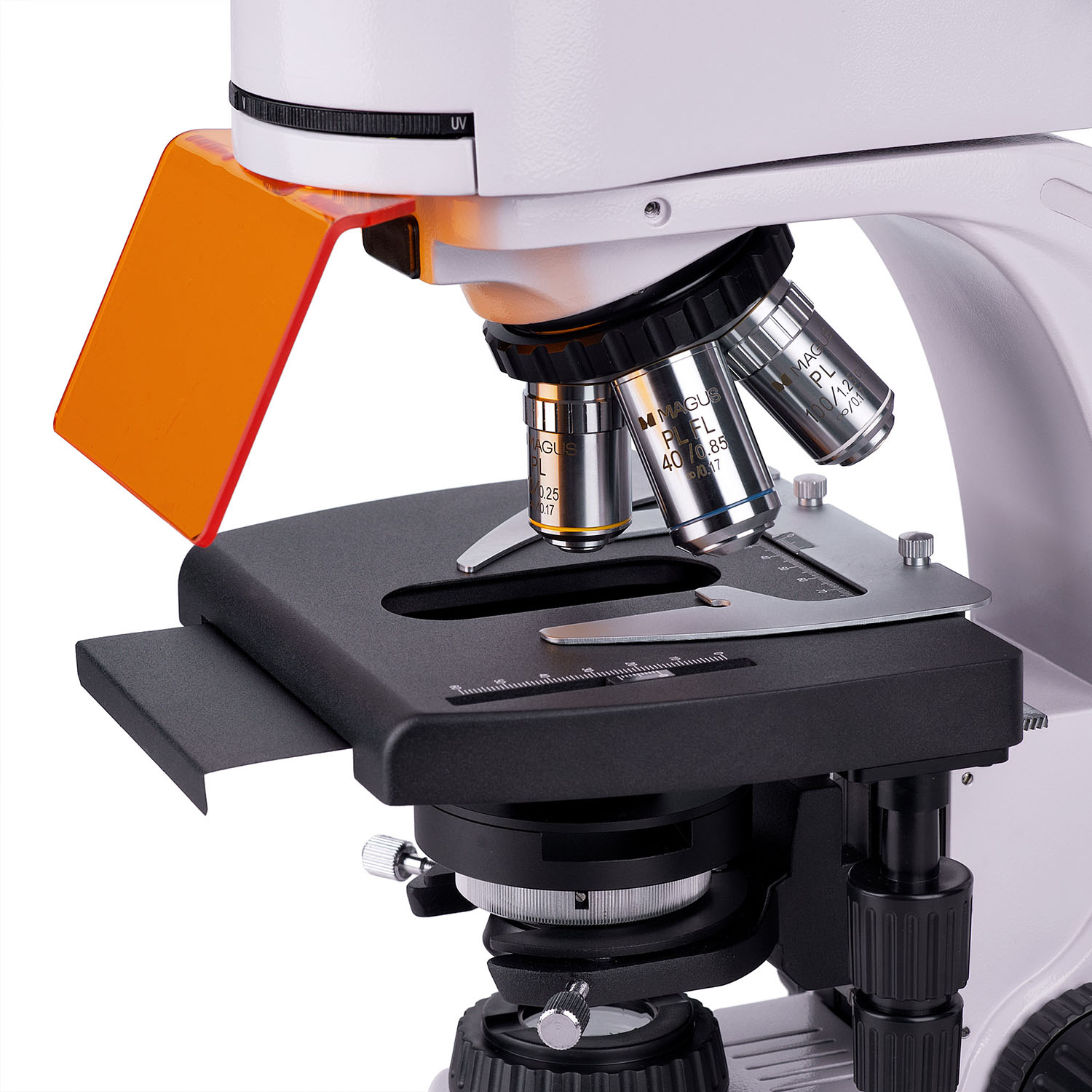
- Infinity plan achromatic objective, fluo: PL FL 40x/0.85 (spring-loaded) WD 0.42mm
- Infinity plan achromatic objective, fluo: PL 100x/1.25 (spring-loaded, oil) WD 0.36mm
- Eyepiece 10x/22mm with long eye relief (2 pcs.)
- UV shield
- C-mount adapter 1x
- Hex key wrench
- Mercury lamphouse power supply
- Power cord
- Reflected light illuminator power cord
- Dust cover
- User manual and warranty card
Available on request:
- 10x/22mm eyepiece with a scale
- 12.5x/14mm eyepiece (2 pcs.)
- 15x/15mm eyepiece (2 pcs.)
- 20x/12mm eyepiece (2 pcs.)
- 25x/9mm eyepiece (2 pcs.)
- Infinity plan achromatic objective, fluo: PL FL 10x/0.35 WD 2.37mm
- Infinity plan achromatic objective: PL 60x/0.80 ∞/0.17 WD 0.46mm
- Phase contrast device
- Darkfield condenser
- Immersion darkfield condenser
- Darkfield slider
- Polarization device
- Calibration slide
Specifications:
| Product ID | 83017 |
| Brand | MAGUS |
| Warranty | 5 years |
| EAN | 5905555018164 |
| Package size (LxWxH) | 17.7x11.8x37.8 cm |
| Shipping Weight | 42.4 kg |
| Type | biological, light/optical, digital |
| Head | trinocular |
| Nozzle | Gemel head (Siedentopf, 360° rotation) |
| Head inclination angle | 30 ° |
| Magnification, x | 40–1000 basic (*optional: 40–1250/1500/2000/2500) |
| Eyepiece tube diameter, in | 1.2 |
| Eyepieces | 10х/22mm, eye relief: 10mm (*optional: 10x/22mm with scale, 12.5x/14; 15x/15; 20x/12; 25x/9) |
| Objectives | infinity plan achromatic and fluo objectives: PL 4x/0.10, PL 10x/0.25, PL FL 40x/0.85, PL 100x/1.25 (oil); parfocal distance 45mm (*optional: PL FL 10x/0.35, PL 60x/0.80 ∞/0.17) |
| Revolving nosepiece | for 5 objectives |
| Working distance, mm | 19.8 (4x); 5.0 (10x); 0.42 (FL 40x); 0.36 (100x); 2.37 (FL 10х); 0.46 (60х) |
| Interpupillary distance, in | 1.9 — 3 |
| Stage, mm | 180x150 |
| Stage moving range, mm | 75/50 |
| Stage features | two-axis mechanical stage, without a positioning rack |
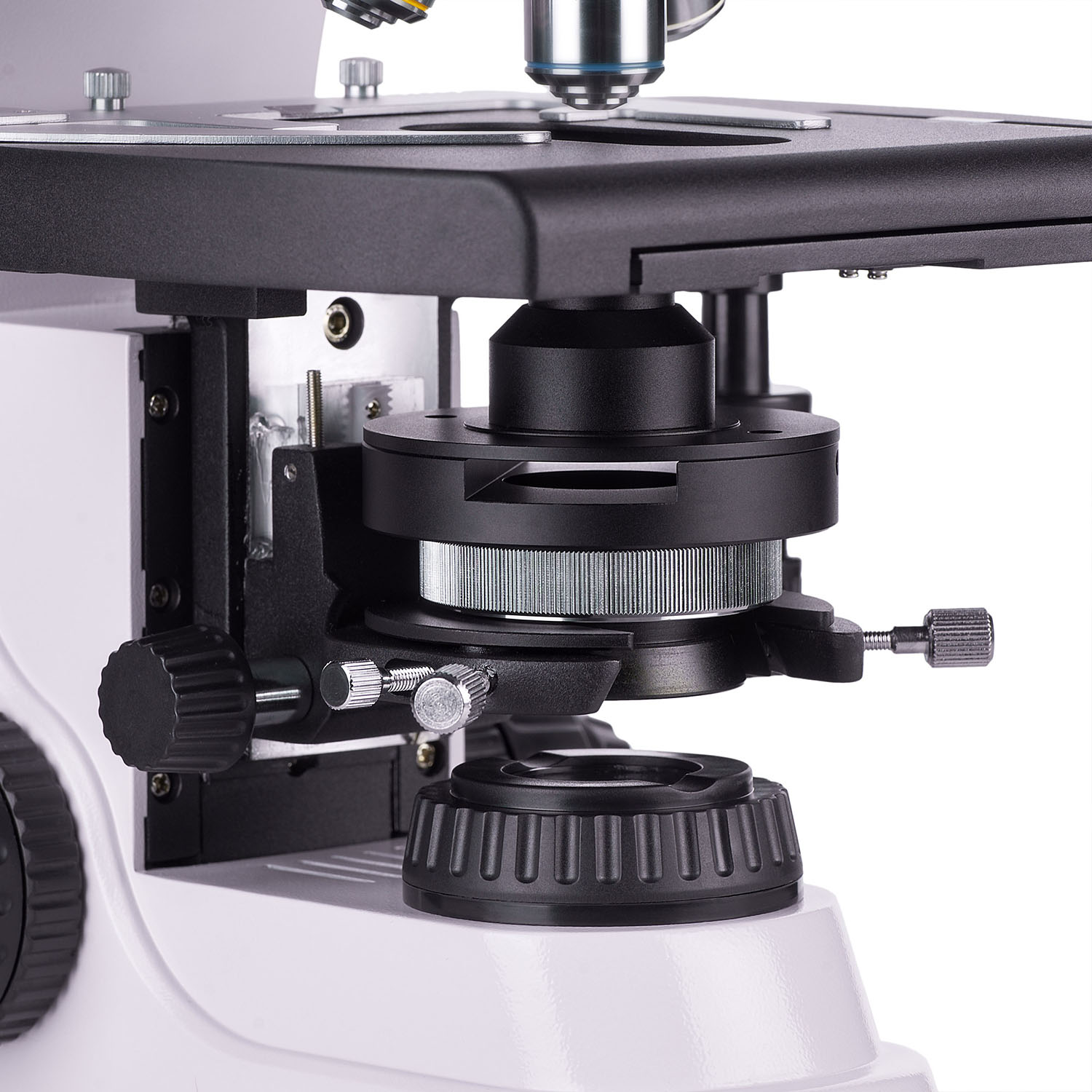
| Condenser | Abbe condenser, N.A. 1.25, center-adjustable, height-adjustable, adjustable aperture diaphragm, a slot for a darkfield slider and phase contrast slider, dovetail mount |
| Diaphragm | adjustable aperture diaphragm, adjustable iris field diaphragm |
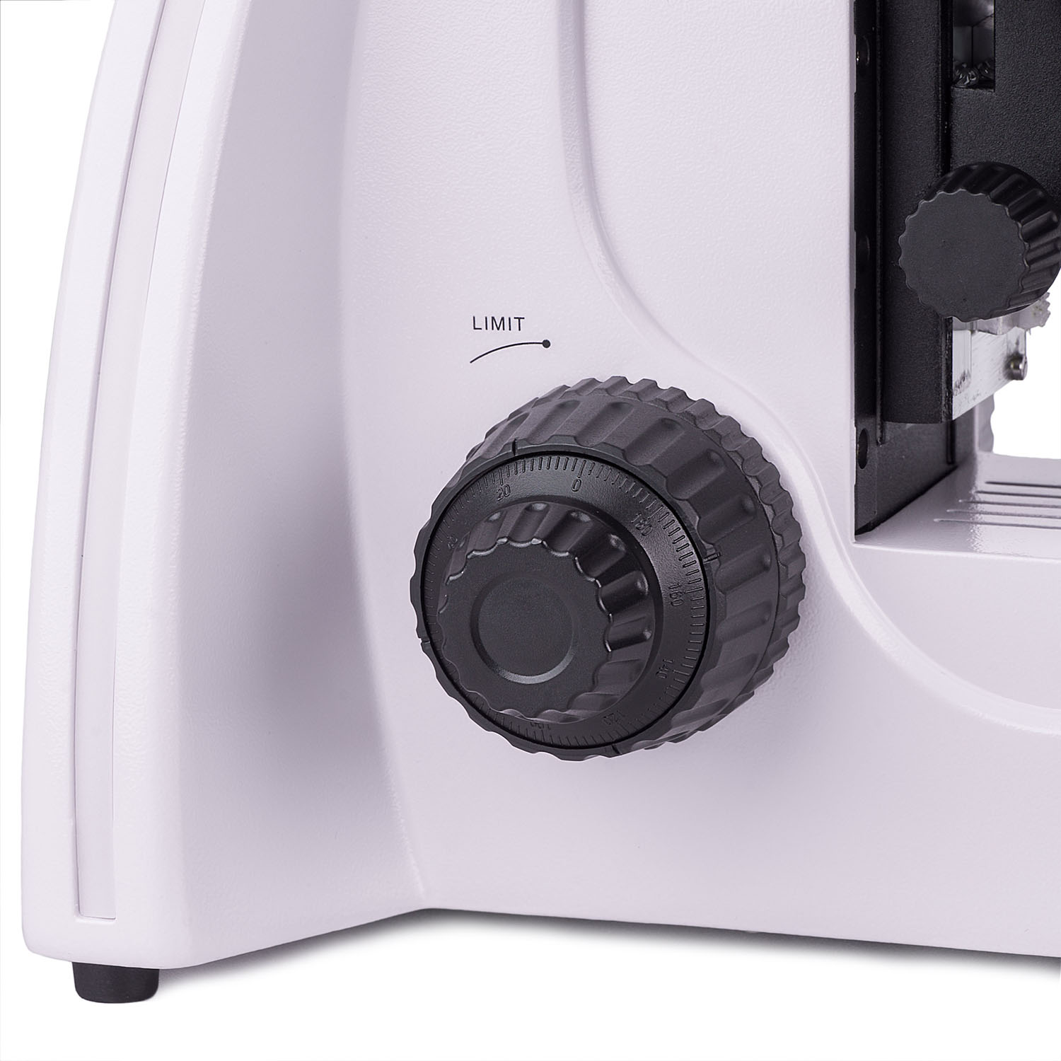
| Focus | coaxial, coarse focusing (21mm, 39.8mm/circle, with a lock knob and tension adjusting knob) and fine focusing (0.002mm) |
| Illumination | fluorescent, halogen |
| Brightness adjustment | ✓ |
| Power supply | AC network, 85–265V, 50/60Hz |
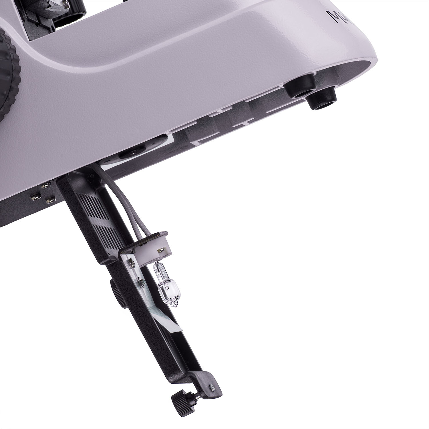
| Light source type | reflected light: 100W mercury lamp; transmitted light: 12V/30W halogen lamp |
| Light filters | yes |
| Operating temperature range, °F | 41...+95 |
| Ability to connect additional equipment | phase contrast device (condenser and objectives), darkfield condenser (dry or oil), polarization devices (polarizer and analyzer), darkfield slider |
| User level | experienced users, professionals |
| Assembly and installation difficulty level | complicated |
| Fluorescent module | filters: ultraviolet (UV), violet (V), blue (B), green (G) |
| Fluorescence filter: filter type, excitation wavelength/dichroic mirror/emission wavelength | ultraviolet (UV), 320–380nm/425 nm/435 nm; violet (V), 380–415nm/455nm/475nm; blue (B), 450–490nm/505nm/515nm; green (G), 495–555nm/585nm/595nm |
| Application | laboratory/medical |
| Illumination location | dual |
| Research method | bright field, fluorescence |
| Pouch/case/bag in set | dust cover |
| Camera specifications | |
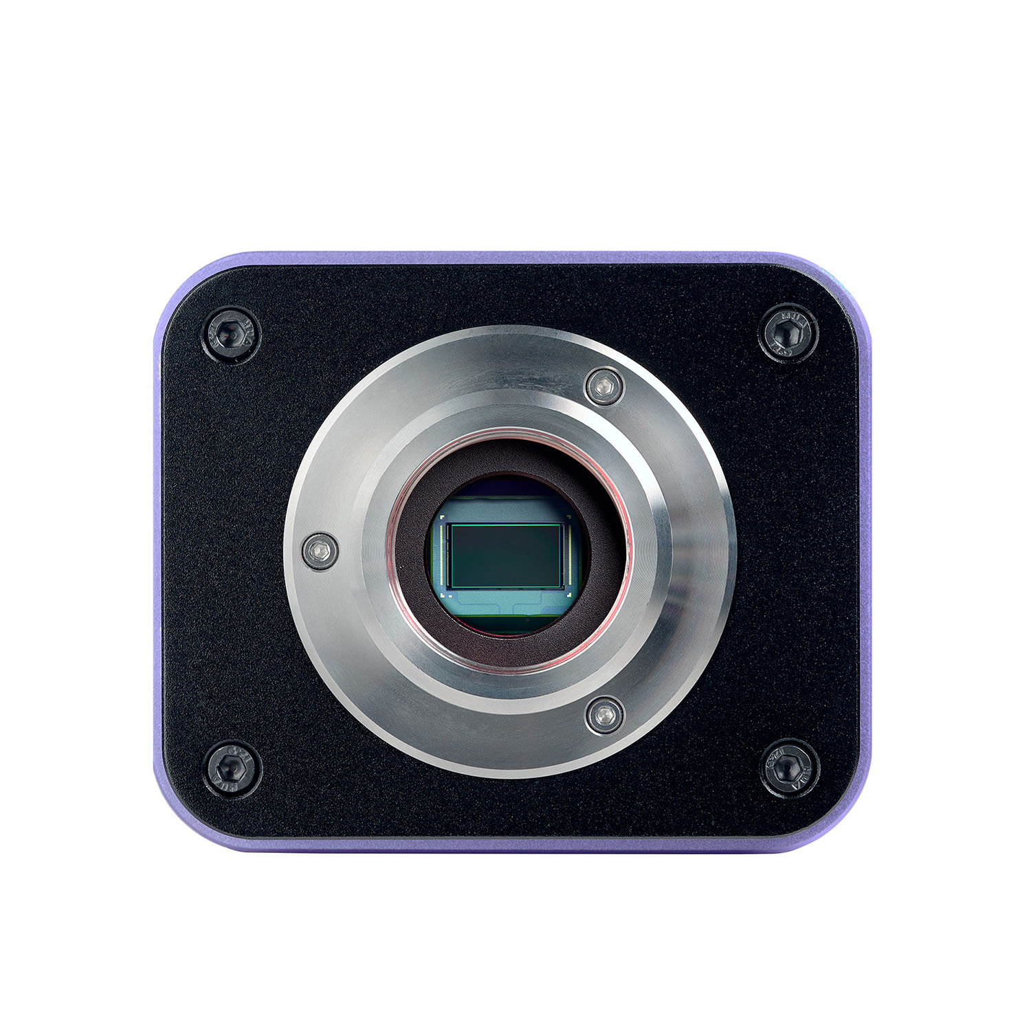
| Sensor | Sony Exmor/Starvis CMOS |
| Color/monochrome | color |
| Megapixels | 8 |
| Maximum resolution, pix | 3840x2160 |
| Sensor size | 1/1.2'' (11.14x6.26mm) |
| Pixel size, μm | 2.9x2.9 |
| Interface connectors | Wi-Fi, HDMI 1.4, USB 3.0 |
| Memory card | SD up to 32GB |
| Ability to connect additional equipment | USB mouse, flash stick (USB), Wi-Fi adapter (USB) |
| Light sensitivity | 1028mV with 1/30s |
| Signal/noise ratio | 0.13mV at 1/30s |
| Exposure time | 0.14ms–1000ms |
| Video recording | ✓ |
| Frame rate, fps at resolution | 30@3840x2160 (HDMI), 30@1920x1080 (Wi-Fi), 30@3840x2160 (USB3.0) |
| Place of installation | trinocular tube, eyepiece tube instead of an eyepiece |
| Image format | *.jpg, *.tif |
| Video format | *.h264/*.h265, *.mp4 |
| Spectral range, nm | 380–650 (built-in IR filter) |
| Shutter type | ERS (electronic rolling shutter) |
| Software | HDMI: built-in; USB: MAGUS View |
| System requirements | Windows 8/10/11 (32bit and 64bit), Mac OS X, Linux, up to 2.8GHz Intel Core 2 or higher, minimum 4GB RAM, USB2.0 port, RJ45, CD-ROM, 19" or larger display |
| Mount type | C-mount |
| Body | CNC aluminum alloy |
| Camera power supply | DC adapter 12V, 1A |
| Camera operating temperature range, °F | -10...+50 |
| Operating humidity range, % | 30 — 80 |
| Monitor specifications | |
| Type of matrix | IPS |
| Display diagonal, inch | 13.3 |
| Display resolution, px | 3840x2160 (4K) |
| Aspect ratio | 16:9 |
| Brightness, cd/m² | 400 |
| Number of displayed colors | 16.7 m |
| Contrast | 1000:1 |
| Horizontal/vertical viewing angle, ° | 178/178 |
| Size of the visible screen area (WxH), mm | 295x165 |
| Pixel pitch (WxH), mm | 0.154x0.154 |
| Frequency of optical source, Hz | 60 |
| Type of matrix backlight | LED |
| LED backlight lifetime, h | 50000 |
| Interface | HDMI |
| Operating temperature range, °F | 5...+131 |
| Operating temperature range, °F | 5...+131 |
| Operating humidity range, % | 10 — 90 |
| Power supply | AC 110–220V, DC 5–12V/1A (Type-C) |
| Power consumption, W | 12 (max.) |
Reviews
Recommended
- On Back Order
0.0 Average Rating Rated (0 Reviews)