Key Features
- High-Resolution Fluorescence & Brightfield: Explore the micro world in stunning detail with 40x-1000x magnification for both fluorescence and brightfield microscopy techniques.
- Capture Images & Videos: The included camera lets you document your discoveries and share them easily.
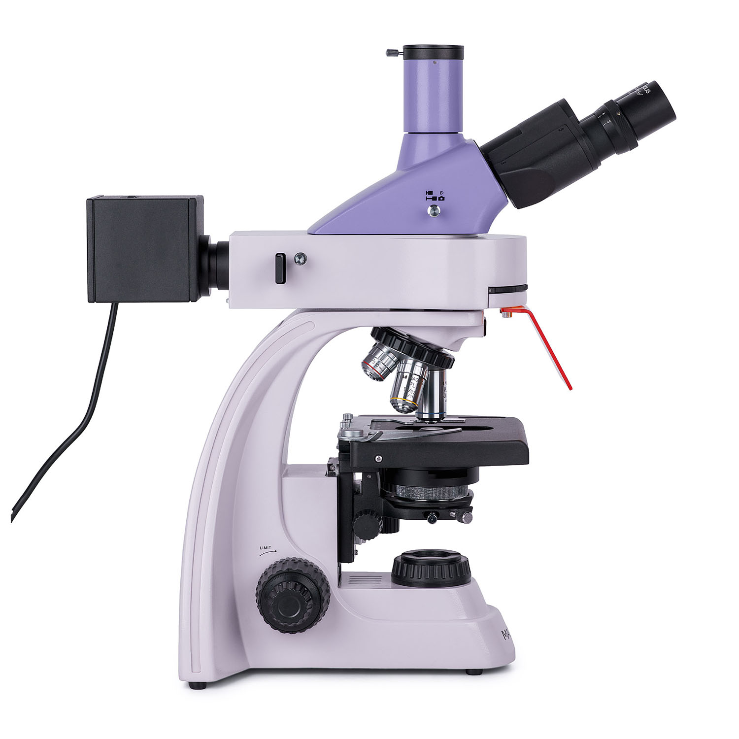
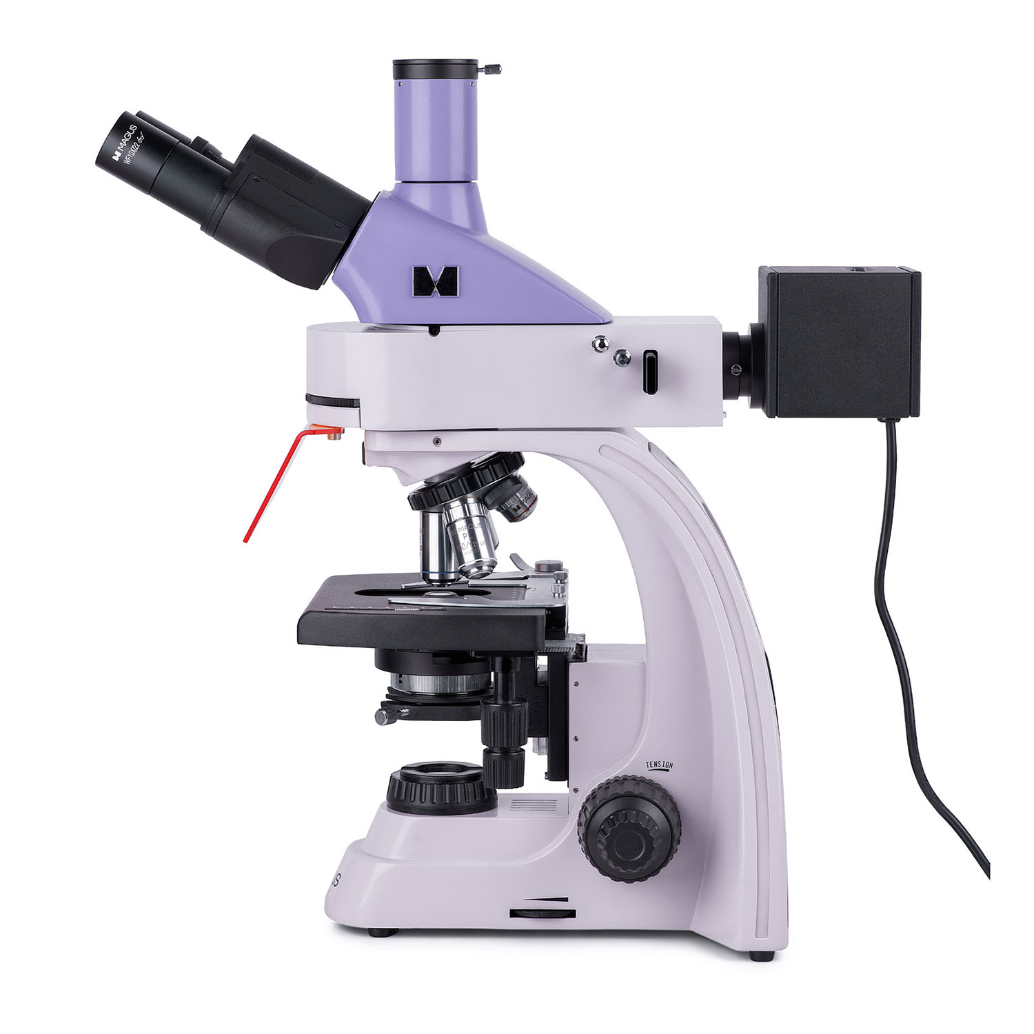
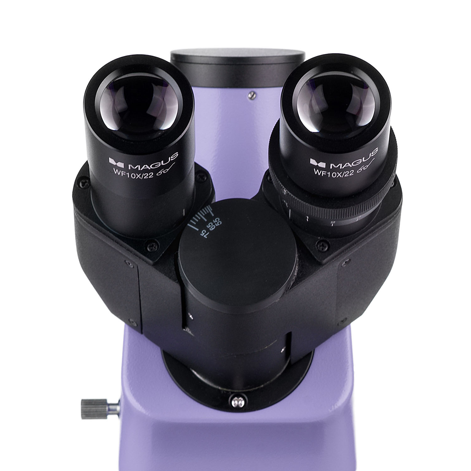
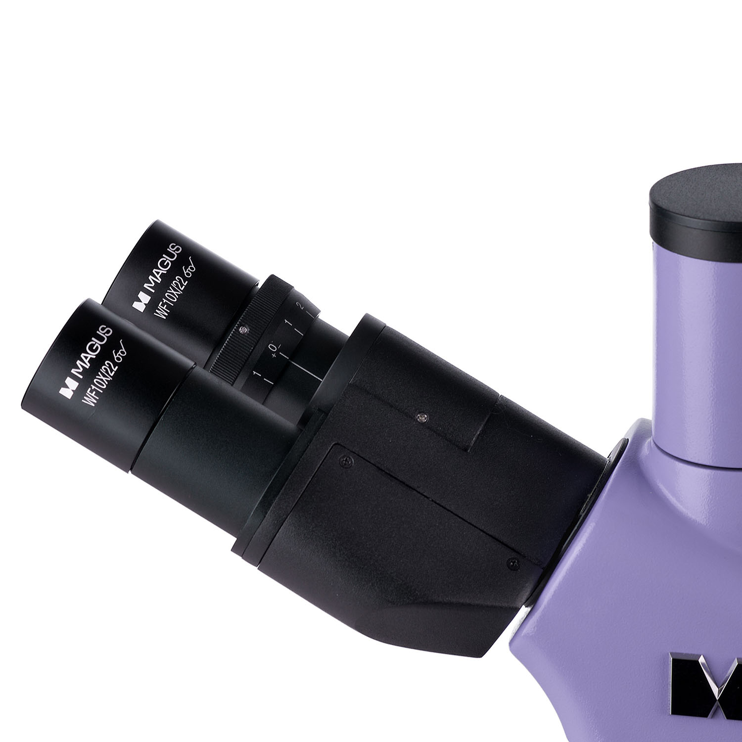
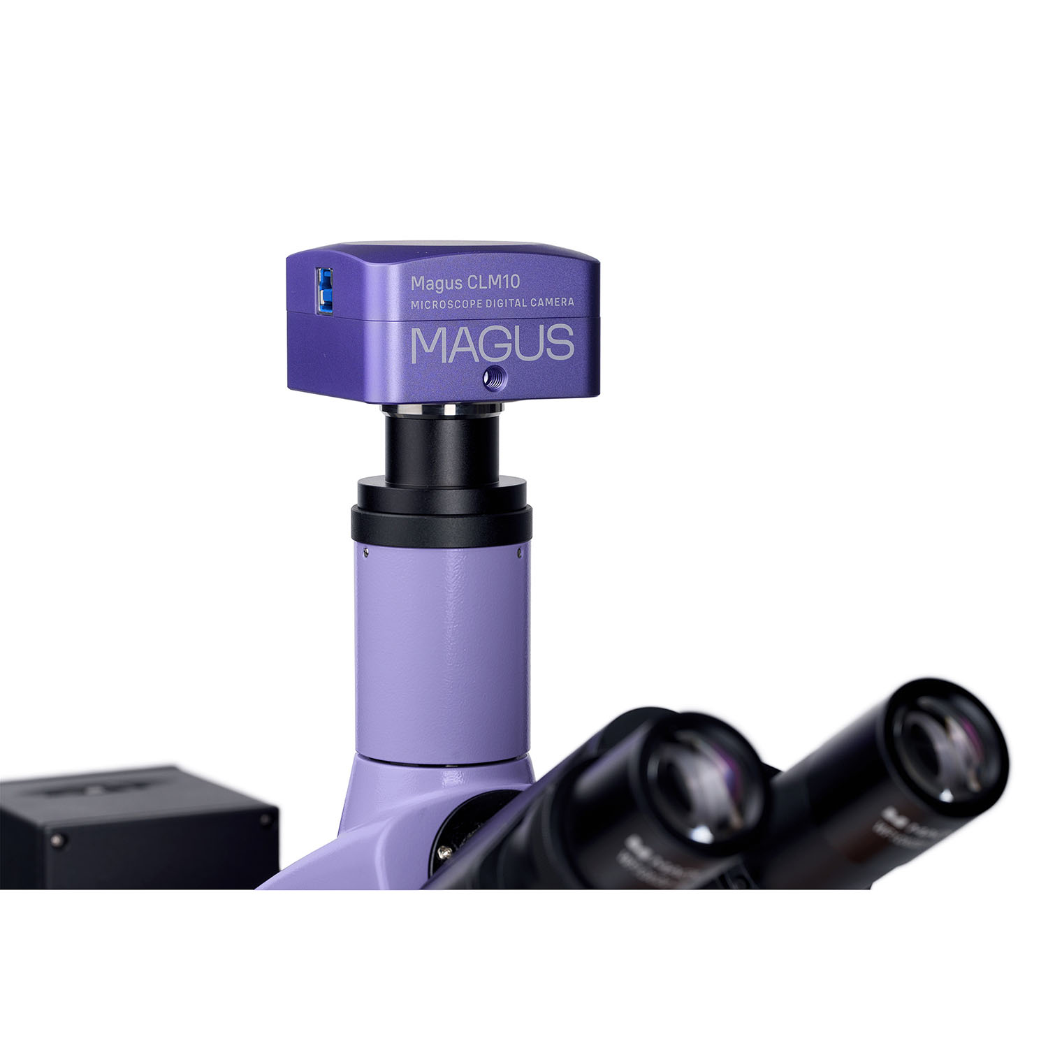
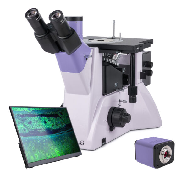
- Trinocular Viewing Head: The trinocular design allows for simultaneous observation through eyepieces and camera attachment for seamless image capture.
- Sharp, Clear Images: Plan achromatic and plan fluorite objectives deliver exceptional image quality and resolution.
- Versatile Illumination: Four reflected light LEDs with different wavelengths and a 3W transmitted LED provide optimal illumination for various samples.
- Köhler Illumination: Ensures even and consistent lighting for superior image clarity in both reflected and transmitted light modes.
Description
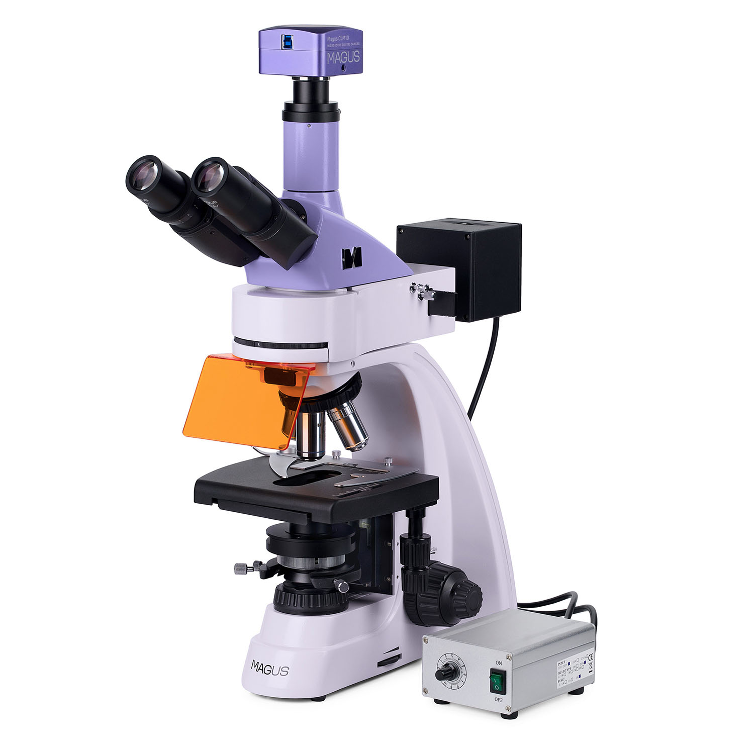
MAGUS Lum D400L: Unveiling the Micro World with Advanced Fluorescence & Brightfield Microscopy
Unleash the Power of Discovery: The MAGUS Lum D400L fluorescence digital microscope empowers researchers, educators, and industrial professionals to delve into the intricacies of the microcosmos. This versatile instrument seamlessly integrates fluorescence and brightfield microscopy techniques, offering a magnification range of 40x to 1000x for detailed exploration of diverse biological, chemical, and material samples.
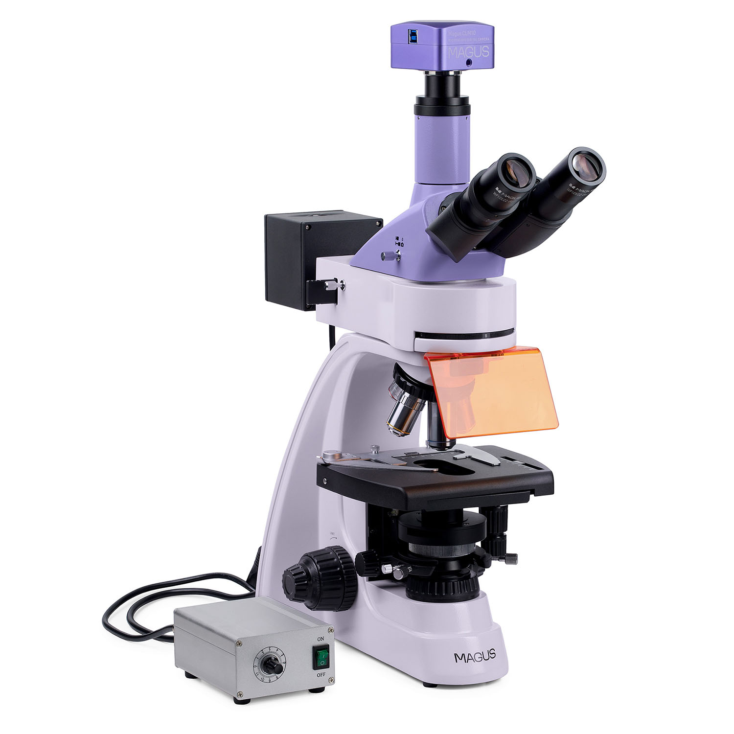
Capture Stunning Visuals: Document your discoveries with exceptional clarity using the included 2.3MP digital camera. The camera boasts a high-resolution output of 1920x1200 pixels, ideal for capturing intricate details. Additionally, the impressive 120fps frame rate enables smooth video recording, perfect for real-time observation and sharing dynamic processes. The USB3.0 interface ensures rapid data transfer, streamlining your workflow.
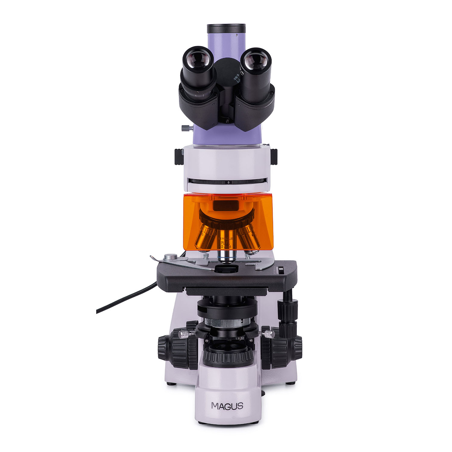
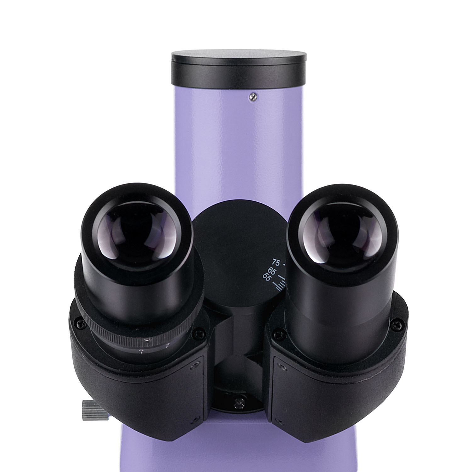
Trinocular Design for Enhanced Collaboration: The trinocular head design of the MAGUS Lum D400L facilitates comfortable visual observation through the eyepieces while simultaneously allowing for camera attachment. This innovative feature promotes seamless collaboration and image sharing, making it ideal for educational and research settings. The 360° rotatable and 30° inclined eyepieces further enhance user comfort during extended observation sessions.
Unmatched Illumination & Clarity: Achieve superior image quality and resolution with Köhler illumination, a technique that ensures even and consistent lighting across the entire field of view. This translates to sharp, high-contrast images for accurate analysis. The microscope utilizes a combination of energy-efficient LED illumination sources: four 5W LEDs for reflected light (offering blue, green, violet, and ultraviolet wavelengths) and a dedicated 3W LED for transmitted light. This versatile illumination system caters to various fluorescence applications and brightfield observations.
Expand Your Microscopy Horizons: The MAGUS Lum D400L is designed for scalability. With additional accessories (sold separately), you can unlock the potential of darkfield, phase contrast, and polarized light microscopy techniques, broadening the range of samples and applications you can explore.
Applications:
- Research & Diagnostics: Medicine, Pharmacology, Forensics, Biotechnology, Veterinary Medicine
- Education & Training: Universities, Colleges, High Schools
- Industrial & Environmental Analysis: Material Science, Quality Control
Who Should Use the MAGUS Lum D400L?
- Researchers seeking a powerful and versatile tool for fluorescence and brightfield microscopy.
- Educators looking for a user-friendly microscope to enhance classroom demonstrations and student engagement.
- Industrial and environmental professionals requiring high-resolution microscopy for material analysis and quality control.
Features:
- Study of samples in reflected light using fluorescence microscopy and in transmitted light using brightfield microscopy
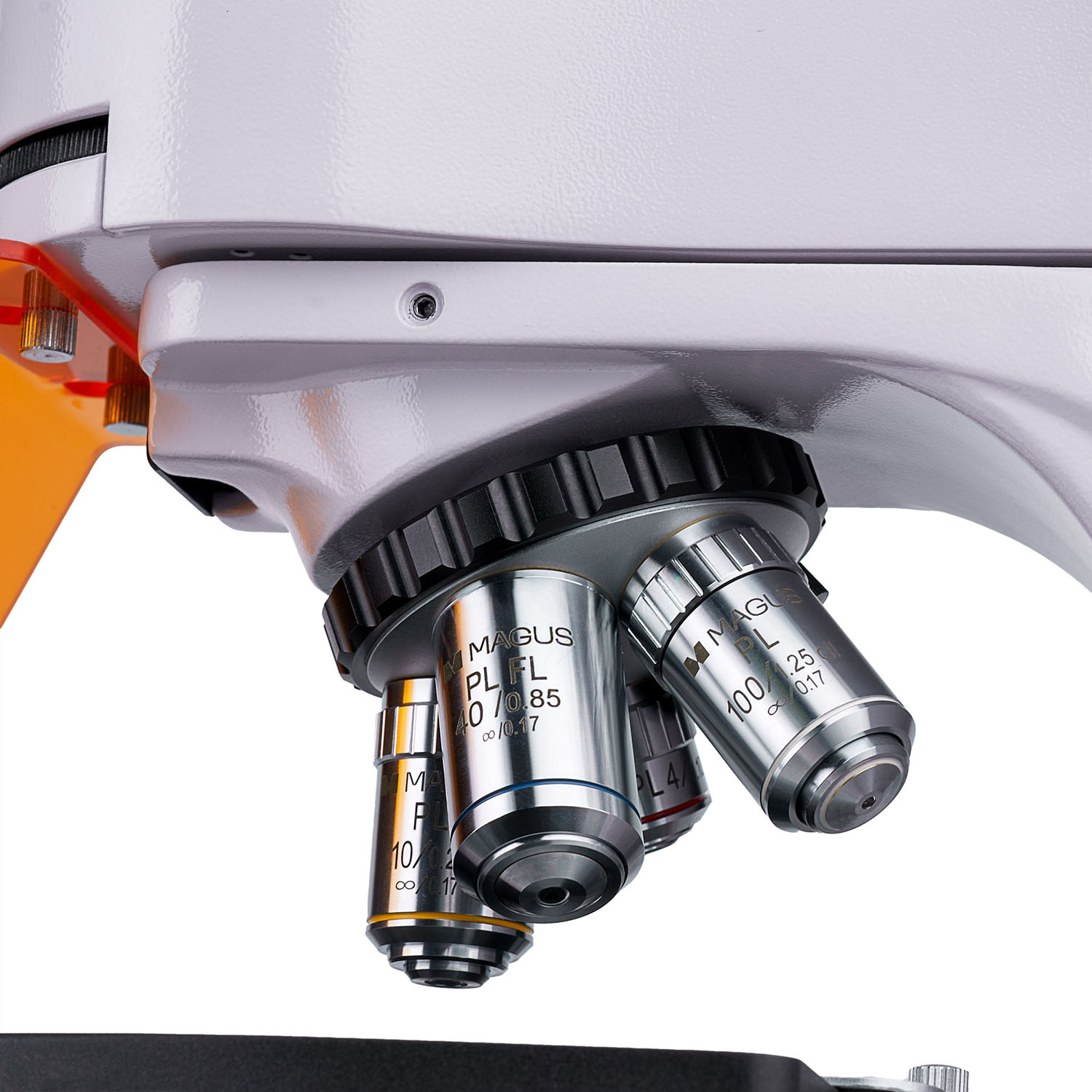
- Infinity plan achromatic objectives: 3 brightfield objectives, 1 fluo objective
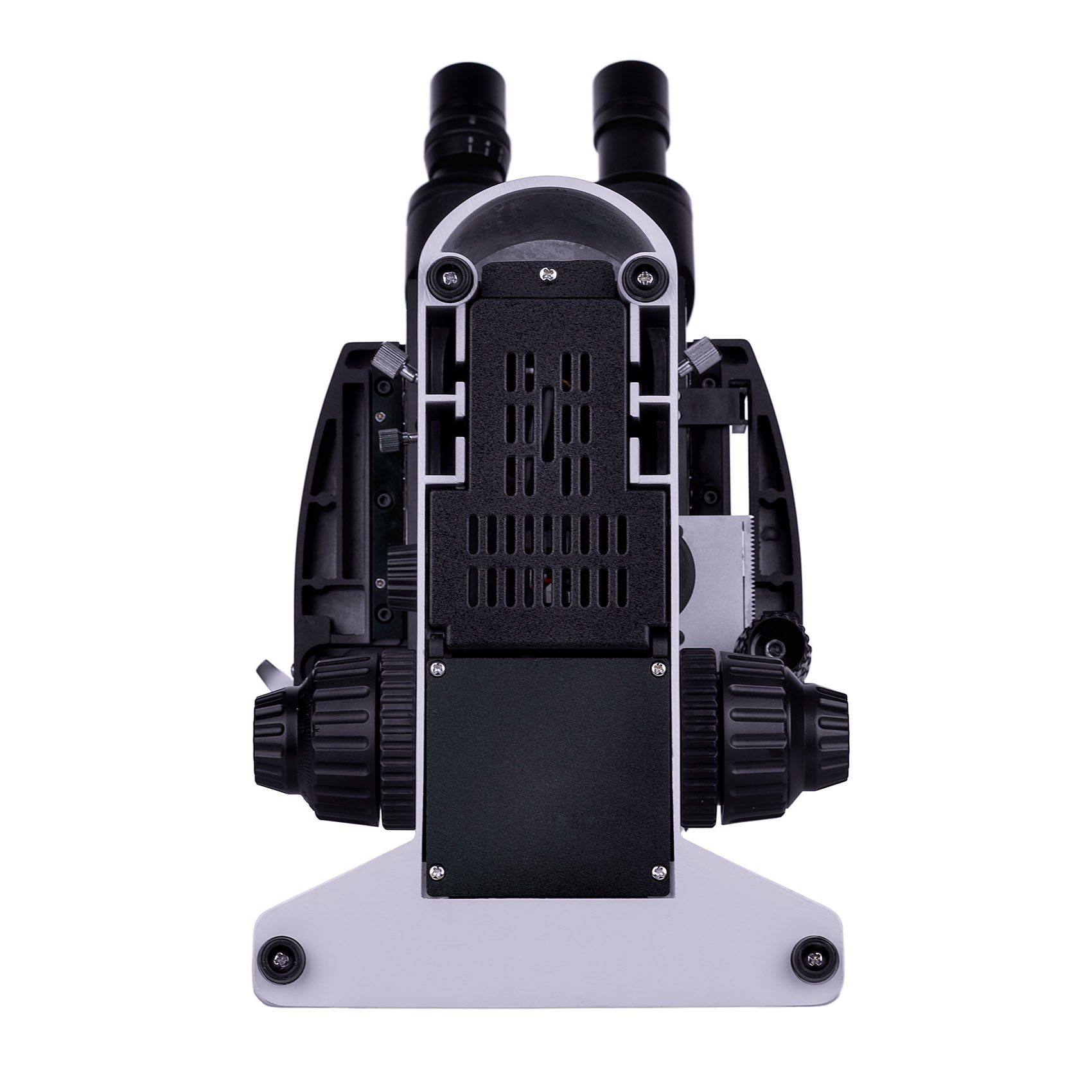
- Trinocular head, 360° rotatable barrels; trinocular tube for mounting a digital camera
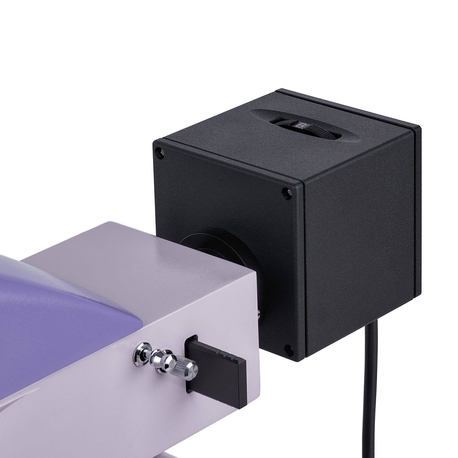
- Four reflected light LEDs operating at different wavelengths: blue (B), green (G), violet (V) and ultraviolet (UV)
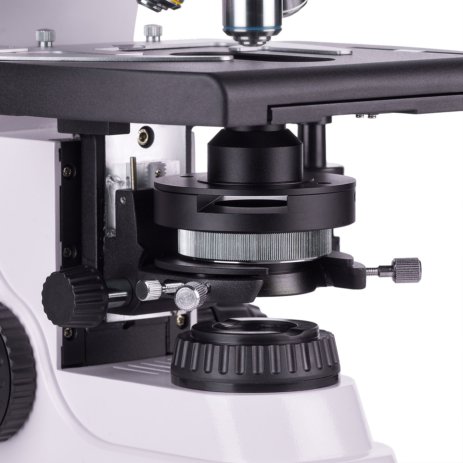
- Transmitted light 3W LED, Köhler transmitted and reflected light illumination
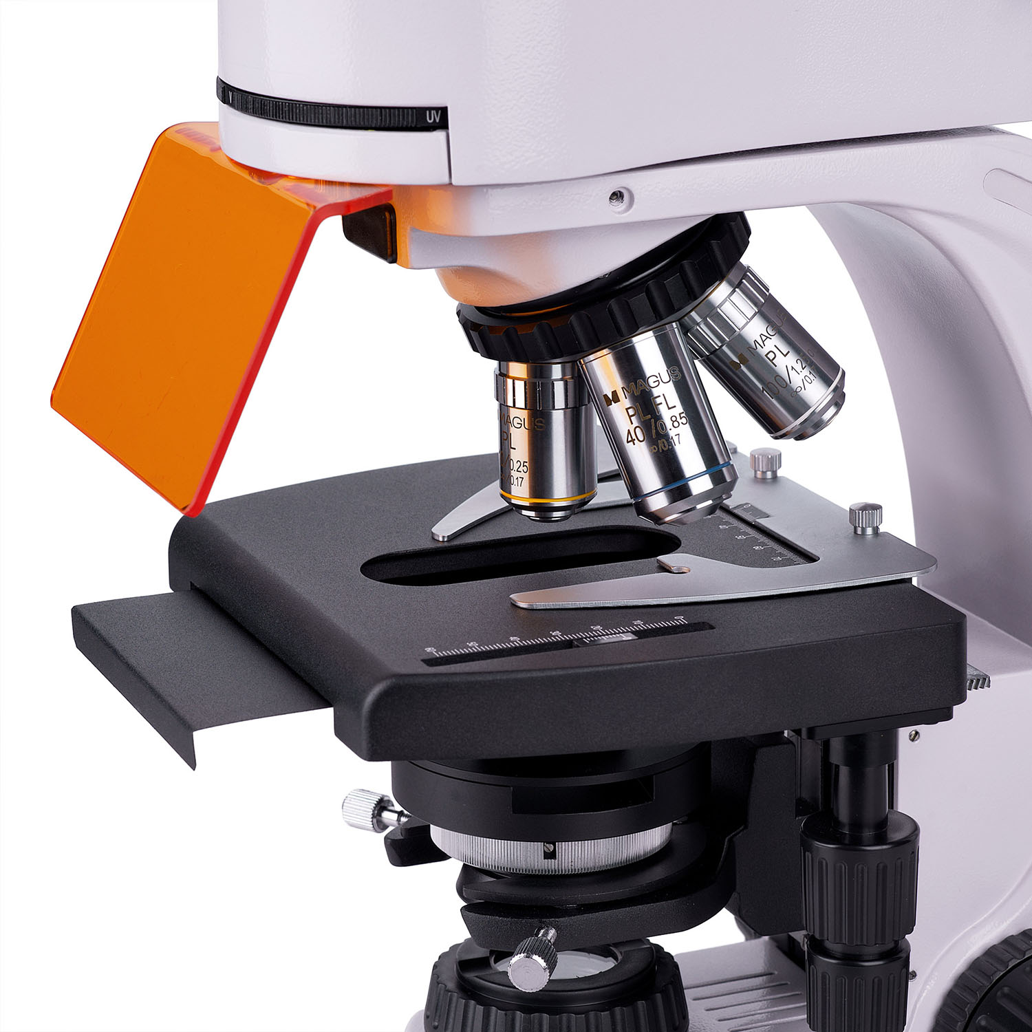
- Stage without a positioning rack, removable specimen holder
- Wide range of accessories (sold separately): eyepieces, objectives, darkfield, polarized light and phase contrast devices
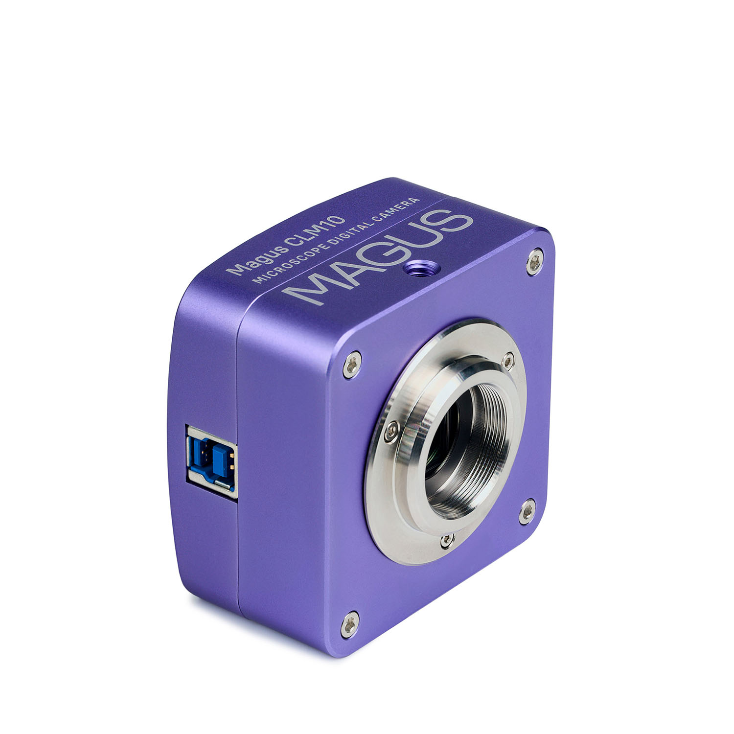
MAGUS CLM10 Digital Camera (Optional):
- Designed for fluorescence and darkfield microscopy with 40x, 60x, and 100x objectives
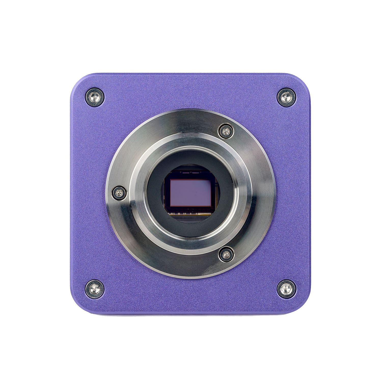
- 2.3MP resolution for detailed images at high magnification; low noise level, low-power dissipation
- 120fps for observing moving objects, recording video, and smooth sample movement
- Global shutter for fast signal readout, increased image brightness, and clear observation of moving objects
- SONY Exmor monochrome CMOS backlit sensor for low noise and high light sensitivity
- USB3.0 interface for fast data transfer
- Software with photo, video recording, editing, external display functions, linear and angular measurements
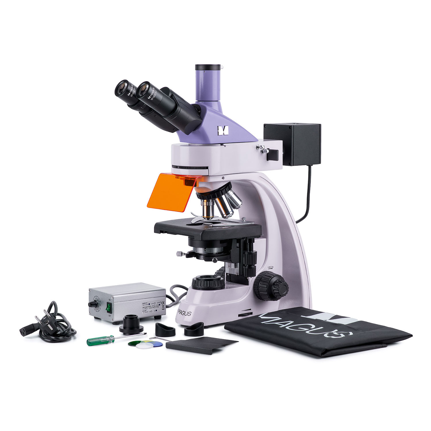
The kit includes:
- MAGUS CLM10 Digital Camera (digital camera, USB cable, installation CD with drivers and software, user manual and warranty card)
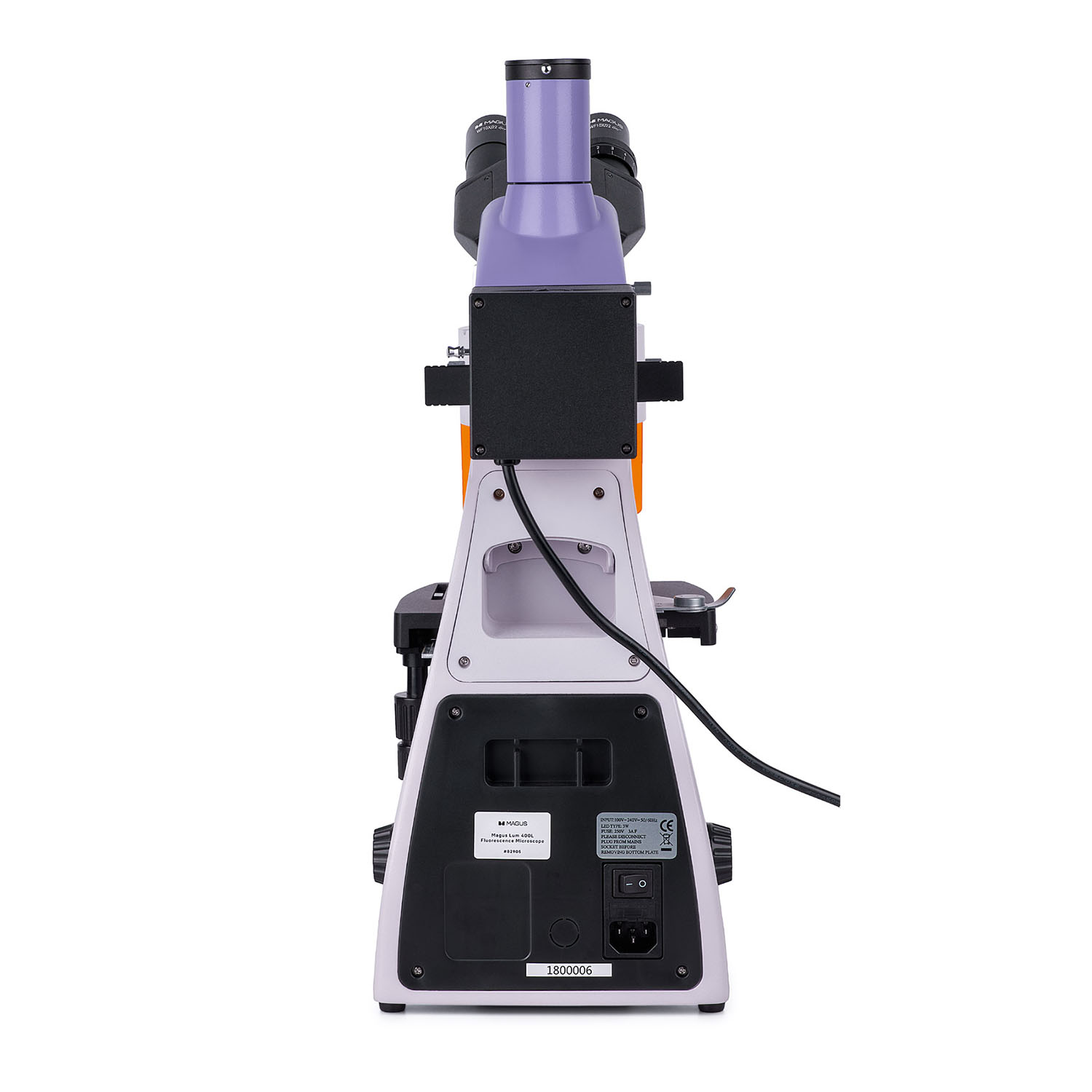
- Base with a power input, transmitted light source and condenser, focusing mechanism, stage, and revolving nosepiece
- Reflected light illuminator
- Lamphouse with LEDs
- Trinocular head
- Infinity plan achromatic objective: PL 4x/0.10 WD 19.8mm
- Infinity plan achromatic objective: PL 10x/0.25 WD 5.0mm
- Infinity plan achromatic objective, fluo: PL FL 40x/0.85 (spring-loaded) WD 0.42mm
- Infinity plan achromatic objective, fluo: PL 100x/1.25 (spring-loaded, oil) WD 0.36mm
- Eyepiece 10x/22mm with long eye relief (2 pcs.)
- UV shield
- C-mount adapter 1x
- Hex key wrench
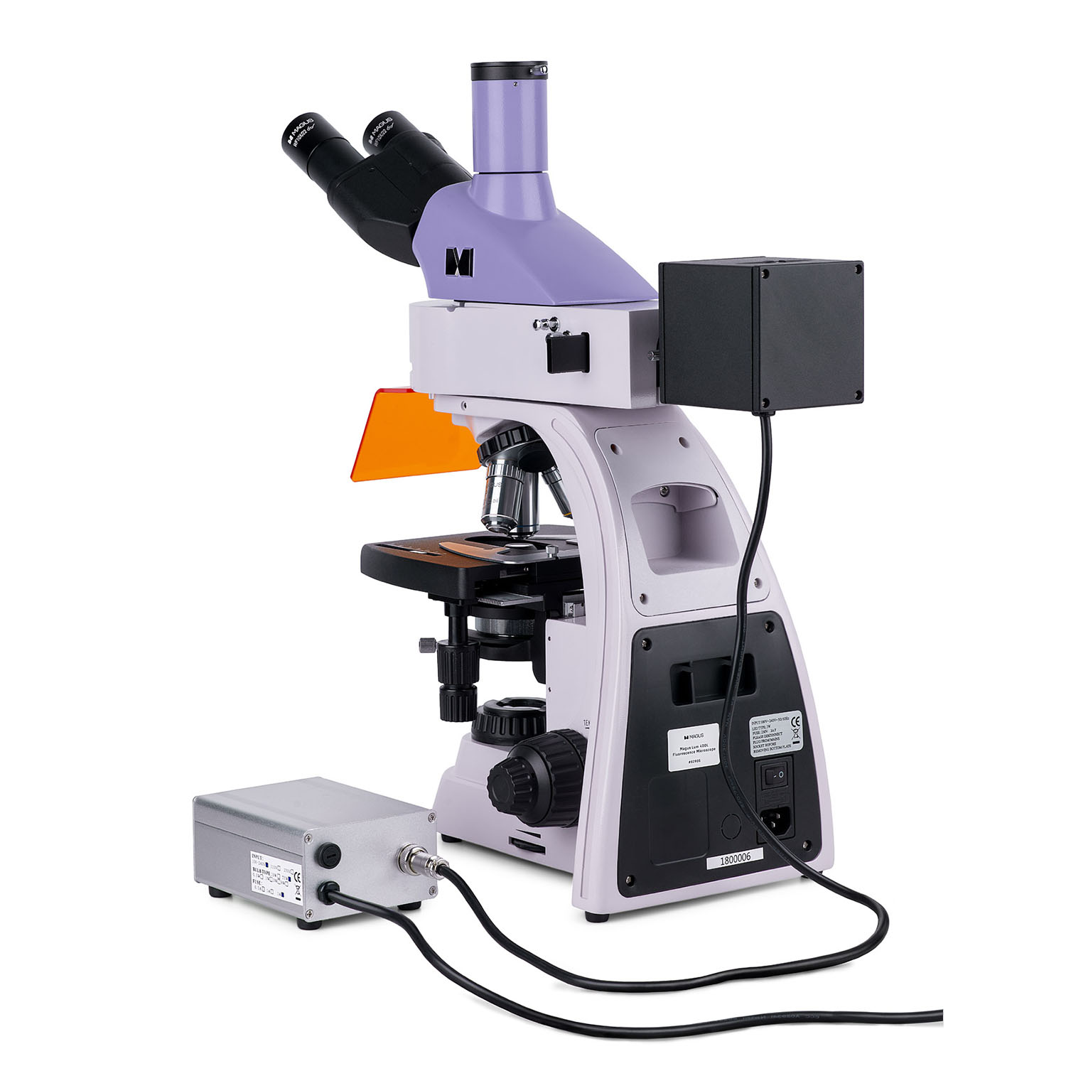
- Reflected-light LEDs power supply
- Power cord
- Reflected light illuminator power cord
- Dust cover
- User manual and warranty card
Available on request:
- 10x/22mm eyepiece with a scale
- 12.5x/14mm eyepiece (2 pcs.)
- 15x/15mm eyepiece (2 pcs.)
- 20x/12mm eyepiece (2 pcs.)
- 25x/9mm eyepiece (2 pcs.)
- Infinity plan achromatic objective, fluo: PL FL 10x/0.35WD 2.37mm
- Infinity plan achromatic objective: PL 60x/0.80 ∞/0.17 WD 0.46mm
- Phase contrast device
- Darkfield condenser
- Immersion darkfield condenser
- Darkfield slider
- Polarization device
- Calibration slide
| Product ID | 83018 |
| Brand | MAGUS |
| Warranty | 5 years |
| EAN | 5905555018188 |
| Package size (LxWxH) | 17.7x11.8x37.8 cm |
| Shipping Weight | 33.3 kg |
| Microscope specifications | |
| Type | biological, light/optical, digital |
| Head | trinocular |
| Nozzle | Gemel head (Siedentopf, 360° rotation) |
| Head inclination angle | 30 ° |
| Eyepiece tube diameter, in | 1.2 |
| Eyepieces | 10х/22mm, eye relief: 10mm (*optional: 10x/22mm with scale, 12.5x/14; 15x/15; 20x/12; 25x/9) |
| Objectives | infinity plan achromatic and fluo objectives: PL 4x/0.10, PL 10x/0.25, PL FL 40x/0.85, PL 100x/1.25 (oil); parfocal distance 45mm (*optional: PL FL 10x/0.35, PL 60x/0.80 ∞/0.17) |
| Revolving nosepiece | for 5 objectives |
| Working distance, mm | 19.8 (4x); 5.0 (10x); 0.42 (FL 40x); 0.36 (100x); 2.37 (FL 10х); 0.46 (60х) |
| Interpupillary distance, in | 1.9 — 3 |
| Stage, mm | 180x150 |
| Stage moving range, mm | 75/50 |
| Stage features | two-axis mechanical stage, without a positioning rack |
| Condenser | Abbe condenser, N.A. 1.25, center-adjustable, height-adjustable, adjustable aperture diaphragm, a slot for a darkfield slider and phase contrast slider, dovetail mount |
| Diaphragm | adjustable aperture diaphragm, adjustable iris field diaphragm |
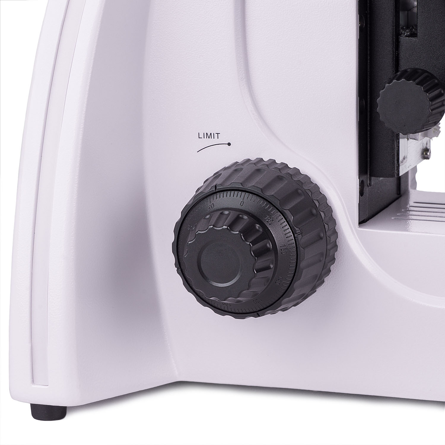
| Focus | coaxial, coarse focusing (21mm, 39.8mm/circle, with a lock knob and tension adjusting knob) and fine focusing (0.002mm) |
| Illumination | LED, fluorescent |
| Brightness adjustment | ✓ |
| Power supply | 85–265V, 50/60Hz |
| Light source type | reflected light: 4 LEDs of different wavelengths, 5W; transmitted light: 3W LED |
| Light filters | yes |
| Ability to connect additional equipment | phase contrast device (condenser and objectives), darkfield condenser (dry or oil), polarization devices (polarizer and analyzer), darkfield slider |
| User level | experienced users, professionals |
| Assembly and installation difficulty level | complicated |
| Fluorescent module | filters: ultraviolet (UV), violet (V), blue (B), green (G) |
| Fluorescence filter: filter type, excitation wavelength/dichroic mirror/emission wavelength | ultraviolet (UV), 320–380nm/420nm/435nm; violet (V), 380–415 nm/460 nm/475nm; blue (B), 410–490nm/505nm/515nm; green (G), 475–550nm/580nm/595nm |
| Application | laboratory/medical |
| Illumination location | dual |
| Research method | bright field, fluorescence |
| Pouch/case/bag in set | dust cover |
| Camera specifications | |
| Sensor | SONY Exmor CMOS |
| Color/monochrome | monochrome |
| Megapixels | 2.3 |
| Maximum resolution, pix | 1920х1200 |
| Sensor size | 1/1.2" (11.25x7.03 mm) |
| Pixel size, μm | 5.86x5.86 |
| Light sensitivity | 1016mV with 1/30s |
| Signal/noise ratio | 0.15mV at 1/30s |
| Exposure time | 0.244ms–2s |
| Video recording | ✓ |
| Video recording | yes |
| Frame rate, fps at resolution | 120@1920x1200 |
| ADC digit capacity (bit) | 8/12 (selectable) |
| Place of installation | trinocular tube, eyepiece tube instead of an eyepiece |
| Image format | *.jpg, *.bmp, *.png, *.tif |
| Video format | output: *.wmv, *.avi, *.h264 (Windows 8 and later), *h265 (Windows 10 and later) |
| Spectral range, nm | 380–650 (with IR filter and anti-reflective filter) |
| Shutter type | Global shutter |
| White balance | manual, automatic |
| Exposure control | manual, automatic |
| Software features | image size, brightness, exposure time |
| Software | MAGUS View |
| Output | USB 3.0, 5Gb/s |
| System requirements | Windows 8/10/11 (32bit and 64bit), Mac OS X, Linux, up to 2.8GHz Intel Core 2 or higher, minimum 2GB RAM, USB3.0 port, CD-ROM, 17" or larger display |
| Mount type | C-mount |
| Body | CNC aluminum alloy |
| Camera power supply | DC, 5V, from computer USB port; a 12V, 3A adapter for Peltier element |
| Camera operating temperature range, °F | -10...+50 |
| Operating humidity range, % | 30 — 80 |
Reviews
Recommended
- On Back Order

0.0 Average Rating Rated (0 Reviews)