


Key Features
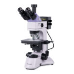
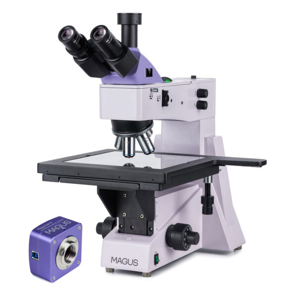
MAGUS Bio D250T Biological Digital Microscope and Camera Features
Key Features of the Microscope
- Trinocular Head: Equipped with a vertical tube for easy digital camera mounting.
- 360° Rotating Eyepiece Tubes: Allows for adjustment to fit the user's height for maximum comfort.
- Diopter Adjustment: Available on the left tube, with coarse focusing including a lock knob and coarse tension adjustment knob, alongside fine focusing.
- Transmitted Light Observations: Utilizes a 30W halogen lamp with adjustable brightness for clear illumination.
- Abbe Condenser: Features an aperture diaphragm and field diaphragm for precise Köhler illumination setup.
- Phase Contrast and Darkfield Options: Special slot in the condenser for installing phase contrast and darkfield sliders.
- Wide Range of Accessories: Compatible with various optional accessories to enhance functionality.
Key Features of the Camera
- 2.1'' Backlit CMOS Sensor: High matrix sensitivity for detailed imaging.
- Versatile Objective Compatibility: Suitable for darkfield or brightfield studies with 40x, 60x, and 100x objectives.
- Flexible Installation: Can be installed into the trinocular tube or eyepiece tube of a microscope.
- Full HD Resolution: Captures images at 1920x1080px with a frame rate of 96fps, ideal for studying moving objects.
- High-Speed Data Transfer: Connects to a PC via USB3.0 port with a data transfer speed of 5Gbps.
- Comprehensive Software Package: Includes necessary software and drivers for seamless integration.
Description
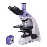
MAGUS Bio D250T Biological Digital Microscope and Camera Description
Description
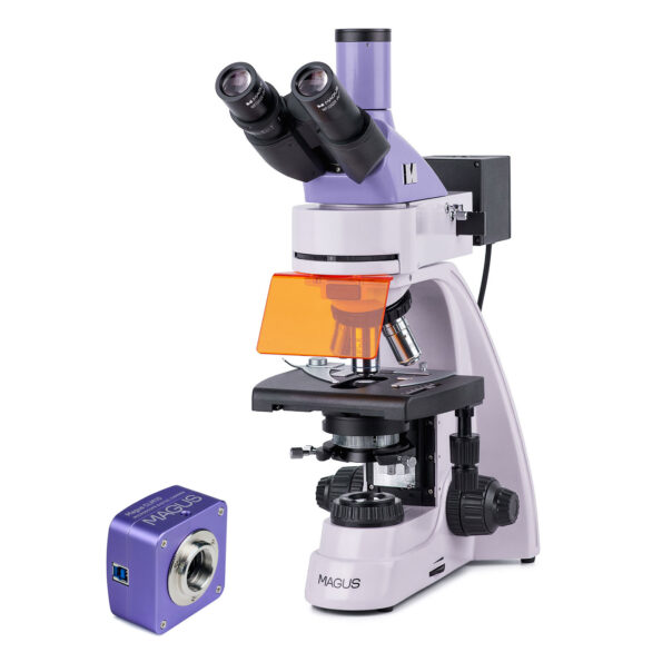
The MAGUS Bio D250T Biological Microscope is a high-end instrument designed for professional observation of biological specimens in transmitted light. Primarily utilizing brightfield techniques, this microscope can also perform darkfield, polarization, and phase contrast microscopy with the appropriate accessories. Ideal for laboratory and research applications, it is widely used in medicine, pharmaceuticals, forensics, agriculture, and other scientific fields.
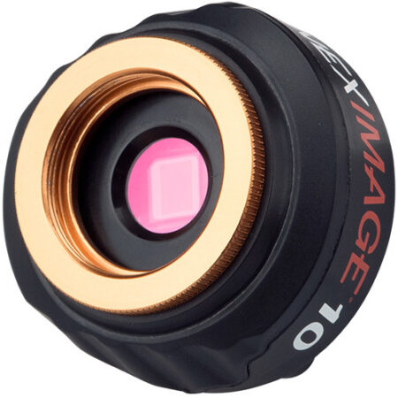
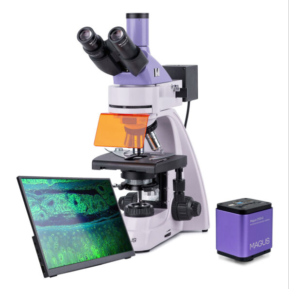
Digital Camera
The full-color camera features a SONY Exmor 2.1MP digital CMOS sensor, using back-illumination technology to minimize light scattering and enhance sensitivity (8935mV at 1/30s). This ensures bright and clear images, even in low light conditions. The camera is optimized for darkfield and brightfield microscopy using objectives with 40x, 60x, and 100x magnifications.
Mounting Options:
- Trinocular Tube: Via a C-mount adapter.
- Eyepiece Tube: Using an adapter and rings.
For the best results, use an adapter with 0.6x to 0.9x magnification to achieve a wide field of view without distortion. The camera connects to a computer through a USB3.0 port, offering a data transfer speed of 5Gbps.
Optics
Plan Achromatic Objectives deliver clear, contrasty images with minimal distortion. The included objectives and eyepieces provide magnifications ranging from 40x to 1000x, expandable with additional eyepieces. The optics are infinity-corrected, allowing for the integration of various accessories into the optical path.
Trinocular Head:
- Vertical Tube: For camera mounting (camera not included).
- 360° Rotating Tubes: Diopter adjustment on the left tube.
- Adjustable Eye Relief: Fits the observer's height.
The revolving nosepiece can hold five objectives simultaneously (four included, one slot free), enabling easy magnification adjustments.
Illumination
A 30W halogen bulb provides bright, warm illumination, which is comfortable for extended use. The brightness is adjustable, and the system is powered by an AC supply.
Köhler Illumination Setup:
- Centered, Height-Adjustable Abbe Condenser
- Aperture Diaphragm
- Field Diaphragm
The condenser includes a slot for darkfield or phase contrast sliders, allowing quick transitions between observation techniques, enhancing performance.
Stage and Focusing Mechanism
The ergonomic stage lacks a positioning rack, enhancing comfort. Objects are moved smoothly with a removable mechanical attachment for manual scanning.
Focusing Mechanism:
- Coaxial Knobs: Located near the base for comfort during long sessions.
- Coarse Focus: Includes a lock knob and tension adjustment.
- Fine Focus: Smooth operation without exertion.
Accessories
The optional accessories include eyepieces, objectives, polarization devices, darkfield condensers, phase contrast devices, calibration slides, and digital cameras. These accessories are specifically designed to optimize the performance of the MAGUS Bio 250T microscope.
The kit contains:
- MAGUS CDF50 Digital Camera (digital camera, USB cable, installation CD with drivers and software, user manual and warranty card)
- Base with a power input, transmitted light source, focusing mechanism, stage, condenser mount, and revolving nosepiece
- Abbe condenser
- Trinocular head
- Infinity plan achromatic objective: 4x/0.10
- Infinity plan achromatic objective: 10x/0.25
- Infinity plan achromatic objective: 40x/0.65 (spring-loaded)
- Infinity plan achromatic objective: 100x/1.25 oil (spring-loaded)
- Eyepiece 10x/22mm with a long eye relief (2 pcs.)
- Eyecup (2 pcs.)
- Filter (4 pcs.)
- C-mount camera adapter
- Bottle of the immersion oil
- AC power cord
- Dust cover
- User manual and warranty card
Available on demand:
- 10x/22mm eyepiece with scale
- 12.5x/14mm eyepiece (2 pcs.)
- 15x/15mm eyepiece (2 pcs.)
- 20x/12mm eyepiece (2 pcs.)
- 25x/9mm eyepiece (2 pcs.)
- Infinity plan achromatic objective: 20x/0.40
- Infinity plan achromatic objective: 60x/0.80 (spring-loaded)
- Phase-contrast device
- Phase slider
- Darkfield condenser NA 0.9
- Oil darkfield condenser NA 1.36–1.25
- Darkfield slider
- Polarization devices
- Calibration slide
- LCD Monitor
specifications:
| Product ID | 83008 |
| Brand | MAGUS |
| Warranty | 5 years |
| EAN | 5905555018027 |
| Package size (LxWxH) | 16.9x10.6x24.8 in |
| Shipping Weight | 24.5 lb |
| Microscope specifications | |
| Type | biological, light/optical, digital |
| Head | trinocular |
| Nozzle | Gemel head (Siedentopf, 360° rotation) |
| Head inclination angle | 30 ° |
| Magnification, x | 40–1000 basic configuration (*optional: 40–1200/1250/1500/1600/2000/2500) |
| Eyepiece tube diameter, in | 1.2 |
| Eyepieces | 10х/22mm, eye relief: 10mm (*optional: 10x/22mm with scale, 12.5x/14; 15x/15; 20x/12; 25x/9) |
| Objectives | infinity plan achromatic: 4x/0.10; 10x/0.25; 40xs/0.65; 100xs/1.25 (oil); parfocal distance: 45mm (*optional: 20x/0.40; 60хs/0.80) |
| Revolving nosepiece | for 5 objectives |
| Working distance, mm | 21 (4x); 5 (10x); 0.66 (40xs); 0.36 (100xs); 8.8 (20x); 0.465 (60xs) |
| Interpupillary distance, in | 1.9 — 3 |
| Stage, mm | 180x150 |
| Stage moving range, mm | 75/50 |
| Stage features | two-axis mechanical stage, without a positioning rack |
| Eyepiece diopter adjustment, diopters | ±5 (on the left tube) |
| Condenser | Abbe condenser, N.A. 1.25, center-adjustable, height-adjustable, adjustable aperture diaphragm, a slot for a darkfield slider and phase contrast slider, dovetail mount |
| Diaphragm | adjustable aperture diaphragm, adjustable iris field diaphragm |
| Focus | coaxial, coarse focusing (21mm, 39.8mm/circle, with a lock knob and tension adjusting knob) and fine focusing (0.002mm) |
| Illumination | halogen |
| Brightness adjustment | ✓ |
| Power supply | 220±22V, 50Hz, AC network |
| Light source type | 12V/30W halogen bulb, G4 |
| Light filters | yes |
| Operating temperature range, °F | 41...+95 |
| Ability to connect additional equipment | phase contrast device (condenser and objectives), darkfield condenser (dry or oil), polarization devices (polarizer and analyzer) |
| User level | experienced users, professionals |
| Assembly and installation difficulty level | complicated |
| Application | laboratory/medical |
| Illumination location | lower |
| Research method | bright field |
| Pouch/case/bag in set | dust cover |
| Weight, lbs | 17.6 |
| Dimensions, in | 200x436x400 |
| Camera specifications | |
| Sensor | SONY Exmor CMOS |
| Sensor size | 1/1.2'' (11.14x6.26mm) |
| Color/monochrome | color |
| Megapixels | 2.1 |
| Maximum resolution | 1920x1080 |
| Pixel size, μm | 5.8x5.8 |
| Light sensitivity | 8935mV with 1/30s |
| Signal/noise ratio | 0.6mV with 1/30s |
| Exposure time | 0.014ms–15s |
| Video recording | yes |
| Frame rate, fps at resolution | 96@1920x1080 |
| Place of installation | trinocular tube, eyepiece tube instead of an eyepiece |
| Image format | *.jpg, *.bmp, *.png, *.tif |
| Video format | output: *.wmv, *.avi, *.h264 (Windows 8 and later), *h265 (Windows 10 and later) |
| Shutter type | ERS (electronic rolling shutter) |
| Spectral range, nm | 380–650 (built-in IR filter) |
| White balance | manual, automatic |
| Exposure control | manual, automatic |
| Software features | image size, brightness, exposure time |
| Output | USB 3.0, 5Gb/s |
| System requirements | Windows 8/10/11 (32bit and 64bit), Mac OS X, Linux, up to 2.8GHz Intel Core 2 or higher, minimum 2GB RAM, USB3.0 port, CD-ROM, 17" or larger display |
| Body | CNC aluminum alloy |
| Software | MAGUS View |
| Camera power supply | DC, 5V, from computer USB port; a 12V, 3A adapter for Peltier element |
| Camera operating temperature range, °F | -10...+50 |
| Operating humidity range, % | 30 — 80 |
Reviews
Recommended
- On Back Order
0.0 Average Rating Rated (0 Reviews)