

The microscopic world is one of the most fascinating areas of modern science. From bacteria and single cells to nanostructures in advanced materials, everything becomes visible through powerful imaging tools. Understanding the Types of microscopes is essential for biology, medicine, material science, and nanotechnology.
In this complete guide, we will explore the full range of Types of microscopes, from basic optical systems to advanced electron and AI-powered technologies. You will also discover real-world applications, global top models, and practical buying guidance.
What Is a Microscope?
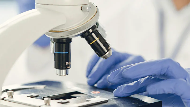
A microscope is a scientific instrument that allows us to observe objects and structures that are too small to be seen with the naked eye. By magnifying tiny specimens, microscopes reveal details that would otherwise remain invisible, helping scientists, researchers, students, and medical professionals better understand the world around us.
From examining bacteria and cells to studying minerals and microelectronic components, microscopes have become essential tools across countless fields of science and industry.
Since the invention of the first microscopes in the 17th century, microscopy technology has advanced dramatically. Early microscopes relied on simple lenses and offered limited magnification, while modern systems can capture highly detailed images at the cellular, molecular, and even atomic levels.
Today, scientists use a wide range of microscope technologies, including optical, digital, electron, and scanning probe microscopes, each designed for specific applications and levels of resolution. These powerful instruments continue to drive discoveries in medicine, biology, materials science, nanotechnology, and many other disciplines.
Main Components of a Microscope
Although microscope designs vary depending on their type and application, most optical microscopes share several essential components that work together to magnify and display a specimen clearly.
- Eyepiece (Ocular Lens): The lens you look through, typically providing 10x magnification.
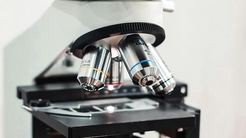
- Objective Lenses: The primary magnifying lenses are positioned close to the specimen, available in different magnification levels such as 4x, 10x, 40x, and 100x.
- Stage: A flat platform that holds the specimen slide securely during observation.
- Stage Clips or Mechanical Stage: Used to keep the slide in place and allow precise movement.
- Illumination System (Light Source): Provides the light needed to view the specimen clearly.
- Condenser: Focuses and directs light onto the sample for improved image quality.
- Iris Diaphragm: Controls the amount of light reaching the specimen to enhance contrast and visibility.
- Coarse Focus Knob: Quickly moves the stage or objective lens for rough focusing.
- Fine Focus Knob: Allows precise focusing for obtaining a sharp and detailed image.
- Revolving Nosepiece: Holds multiple objective lenses and rotates to change magnification levels.
- Arm: Supports the upper parts of the microscope and is commonly used when carrying the instrument.
- Base: The foundation of the microscope that provides stability and often houses the illumination system.
- Body Tube or Head: Connects the eyepiece to the objective lenses and maintains proper optical alignment.
How Does a Microscope Work?
A microscope works by magnifying small objects through a combination of lenses, light, and imaging systems. In a traditional optical microscope, light passes through or reflects from the specimen and is focused by the objective lens to create an enlarged image. This image is then further magnified by the eyepiece, allowing the viewer to observe details that are invisible to the naked eye.
More advanced microscopes use different technologies to achieve higher levels of magnification and resolution. For example, electron microscopes use beams of electrons instead of light, while digital microscopes rely on cameras and computer displays.
Regardless of the technology, the primary purpose of every microscope is the same: to reveal tiny structures and details that help scientists, students, and researchers better understand the microscopic world.
Main Categories of Microscopes
Microscopes are generally divided into three main groups:
- Optical (Light) Microscopes
- Electron Microscopes
- Scanning Probe Microscopes
Each category includes specialized technologies, forming the complete structure of modern Types of microscopes.
1. Optical Microscopes (Light Microscopes)
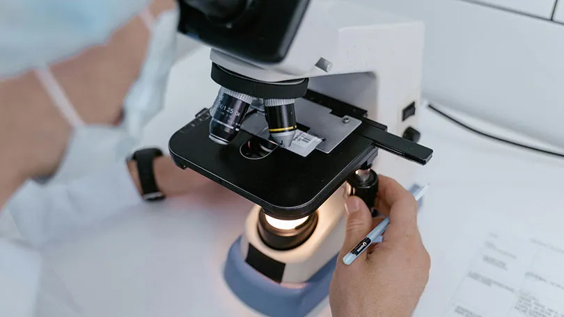
Optical microscopes, also known as light microscopes, are the most widely used microscopes in schools, universities, research laboratories, and medical facilities. They work by using visible light and a system of lenses to magnify specimens, making it possible to observe cells, tissues, microorganisms, and small structures that cannot be seen with the naked eye.
Due to their ease of use, affordability, and versatility, optical microscopes remain the first choice for many educational and scientific applications. Optical microscopes are generally divided into two main categories:
Compound Microscopes
Compound microscopes are designed for viewing thin and transparent specimens at relatively high magnifications. They use multiple lenses, including objective lenses and an eyepiece, to produce detailed images of biological samples.
These microscopes are commonly used in biology, medicine, microbiology, and laboratory research for examining cells, tissues, bacteria, and other microscopic organisms.
Stereo Microscopes
Stereo microscopes, also known as dissecting microscopes, provide a three-dimensional view of a specimen at lower magnifications. Unlike compound microscopes, they are ideal for observing larger and solid objects such as insects, plants, gemstones, electronic components, and mechanical parts. Their 3D visualization makes them particularly useful for inspection, assembly work, and educational demonstrations.
Advanced Optical Microscopy Techniques
Modern optical microscopy includes several specialized imaging methods that improve contrast and reveal details that may not be visible with standard illumination:
- Brightfield Microscopy: The most common technique, ideal for stained biological specimens.
- Phase-Contrast Microscopy: Enhances the visibility of transparent and living cells without staining.
- Fluorescence Microscopy: Uses fluorescent dyes to visualize specific cellular structures, proteins, and microorganisms.
- Darkfield Microscopy: Produces bright images of specimens against a dark background, making fine details easier to observe.
These advanced techniques have significantly expanded the capabilities of optical microscopes, allowing scientists to study living organisms, bacteria, cells, and microscopic structures with greater clarity and precision.
2. Electron Microscopes
Electron microscopes use beams of electrons instead of light, enabling extremely high magnification:
- Transmission Electron Microscope (TEM): Used to study the internal structures of cells and materials at the atomic level.
- Scanning Electron Microscope (SEM): Produces detailed 3D surface images for materials, biology, and industrial applications.
- Types of Electron Microscope (Advanced Systems): Includes Cryo-EM and STEM technologies used in modern nanoscience and virology.
3. Scanning Probe Microscopes
Scanning Probe Microscopes (SPMs) are among the most advanced microscopy techniques, enabling scientists to examine surfaces at the nanometer and atomic scales. Unlike optical and electron microscopes, these systems use an ultra-fine physical probe that moves across the surface of a specimen to collect detailed information about its structure and properties.
The two most common types are the Atomic Force Microscope (AFM), which measures tiny forces between the probe and the sample to create highly detailed three-dimensional surface images, and the Scanning Tunneling Microscope (STM), which uses quantum tunneling principles to visualize and even manipulate individual atoms.
These powerful instruments play a critical role in nanotechnology, materials science, semiconductor development, molecular biology, and advanced physics research.
4. Digital and AI Microscopes
Modern microscopy has evolved beyond traditional optical systems and now includes advanced digital and AI-powered technologies. Digital microscopes use built-in cameras and high-resolution displays to provide real-time imaging, allowing users to view, record, and analyze specimens more easily and efficiently.
In addition, AI-powered systems enhance microscopy by automatically recognizing patterns, detecting abnormalities, and assisting in data analysis, especially in medical and research applications. Automated diagnostic systems further improve speed and accuracy, making these smart microscopes an important part of modern scientific workflows in biology, medicine, and industrial research.
How to Choose the Right Microscope
- Biology: Optical / Fluorescence
- Medicine: Compound / Electron
- Nanotechnology: AFM / STM / TEM
- Industry: SEM / Digital systems
Top 5 Advanced Microscope Systems Worldwide
Modern microscopy has pushed the boundaries of physics, combining electron beams, super-resolution lasers, and AI to visualize everything from individual atoms to live cellular processes. Here are the top 5 advanced microscope systems worldwide:
1. ZEISS Axio Imager 2:
The ZEISS Axio Imager 2 is a top-tier research microscope used in medical and biological labs. It delivers extremely sharp imaging and is widely used in neuroscience, pathology, and cell biology. It is also commonly associated with Biological Microscopes for advanced applications in biological and life science research.
2. Leica DM6 B: The Leica Microsystems DM6 B is a fully automated digital microscope system. It integrates AI tools and advanced imaging, making it ideal for clinical and pharmaceutical research.
3. Nikon Eclipse Ni Series: The Nikon Eclipse Ni series offers excellent optical performance for life science research. It is commonly used in cell biology, microbiology, and medical diagnostics.
4. JEOL JEM-2100 TEM: The JEOL JEM-2100 is a high-end Transmission Electron Microscope used for atomic-level imaging. It is essential in nanotechnology, virology, and materials science.
5. Thermo Scientific Helios 5 CX: The Thermo Fisher Scientific Helios 5 CX combines SEM and FIB technology. It is widely used in semiconductor research, nanofabrication, and advanced material analysis.

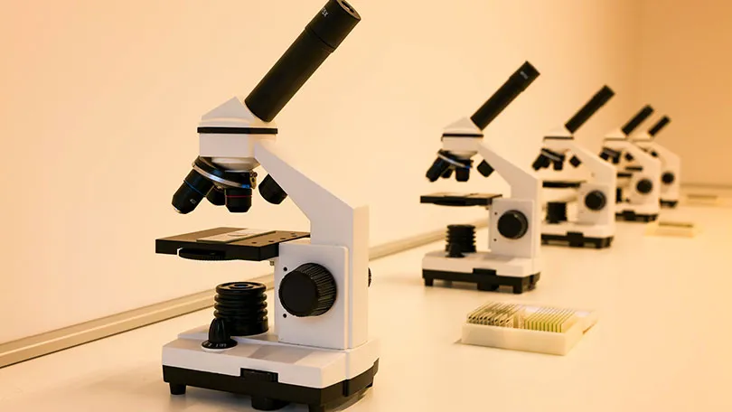
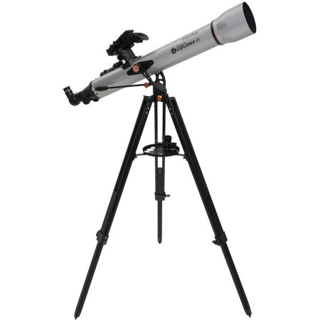
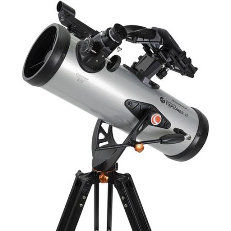
The Best Microscopes from Our Collection
With over 25 years of experience in Iran and 15 years of active presence in Dubai, we have established a trusted name in the field of optical instruments and scientific equipment. In Iran, we operate through dubaitelescope.com, while in Dubai, we are known as Magic Shop, serving customers with reliable products and professional support.
You can also browse our collection of high-quality microscopes available directly through our store and choose the model that best fits your needs, whether for education, research, or professional use, including Metallurgical Microscopes.
- Biological Compound Microscopes: Perfect for students and laboratories studying cells and bacteria.
- Digital USB Microscopes: Ideal for electronics inspection and real-time computer imaging.
- Fluorescence Microscopes: Used in advanced biology for DNA and protein research.
- Stereo Microscopes: Great for industrial inspection and repair work.
- Advanced Research Microscopes: Suitable for universities and nanotechnology laboratories.
Why Understanding Types of Microscope Matters
Understanding the Types of microscopes is essential because each microscope is designed for a specific purpose and level of detail. Choosing the right type directly affects the accuracy and quality of results, whether in education, medical diagnosis, industrial inspection, or advanced scientific research.
From simple optical microscopes used in classrooms to powerful electron and scanning probe systems capable of observing atoms, each tool provides a different level of magnification and insight. Knowing these differences helps users select the most suitable microscope for their needs and ensures more precise, reliable, and meaningful observations.
The Future of Microscopy
The future of microscopy is rapidly evolving with the integration of advanced technologies that are transforming how we study the microscopic world. Modern systems are increasingly moving toward AI-assisted diagnosis, enabling faster and more accurate detection of diseases and abnormalities.
At the same time, innovations such as 3D cellular imaging are allowing scientists to observe cells in more realistic and detailed structures than ever before.
Fully automated research systems are improving efficiency in laboratories by reducing manual work and increasing consistency in results, while nano-level precision analysis is pushing the limits of what we can observe at atomic and molecular scales. Together, these advancements are reshaping both science and medicine, opening new possibilities for discovery and innovation.
Conclusion
Microscopy has become one of the most important tools in modern science, allowing us to explore a world that is completely invisible to the naked eye. From basic optical systems to advanced electron, scanning probe, digital, and AI-powered technologies, each type of microscope plays a unique role in expanding our understanding of life and matter.
With continuous technological progress, microscopy is becoming more precise, faster, and more intelligent, opening new doors in medicine, biology, nanotechnology, and industry.
Ultimately, understanding the Types of microscopes helps us choose the right tool for the right task and brings us closer to uncovering the smallest secrets of the universe.
FAQ
Optical, electron, scanning probe, and digital microscopes.
Compound and fluorescence microscopes are best for cell observation.
Electron microscopes like TEM offer atomic-level resolution.
They allow scientists to study invisible structures like cells, bacteria, and atoms.


No Comments