
For most of human history, scientific observation depended entirely on the human eye.
Early astronomers stared through primitive telescopes and sketched what they saw by hand. Naturalists observed insects, plants, and microorganisms through simple lenses and carefully recorded details in notebooks. Even after the invention of the microscope in the 17th century, microscopic observation remained deeply personal and subjective. What one observer saw depended on eyesight, experience, lighting conditions, and artistic skill. The microscope opened hidden worlds, but those worlds could not easily be shared. A scientist observing bacteria in one country could only describe the observation through words or drawings. Another observer elsewhere might interpret the same specimen differently. In many ways, microscopy was limited not only by optics, but also by human perception itself. Digital microscopy changed this forever.
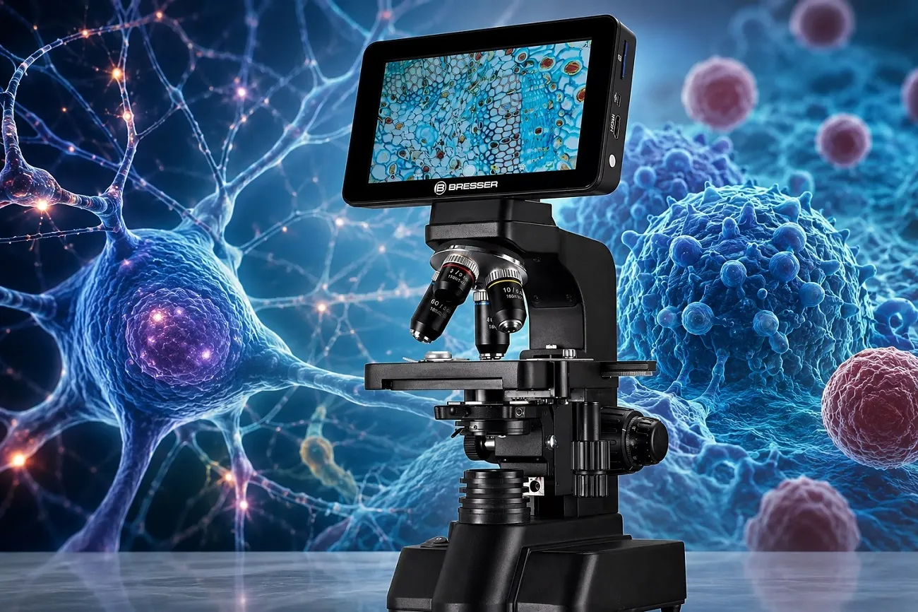
Today, microscopic images can be captured instantly, displayed on giant monitors, analyzed by software, transmitted across continents, enhanced by artificial intelligence, measured automatically, and archived permanently. A modern microscope camera transforms the microscope from a purely visual instrument into a digital imaging system capable of communication, documentation, automation, and analysis.
The rise of digital microscopy represents one of the most important changes in the history of scientific observation. What began as a simple effort to attach cameras to microscopes has evolved into an entire technological ecosystem involving:
- CMOS sensors,
- scientific imaging,
- real-time measurement,
- image stacking,
- fluorescence imaging,
- machine learning,
- AI-assisted diagnostics,
- and remote scientific collaboration.
Today, microscope cameras are used not only in laboratories, but also in:
- education,
- medicine,
- industry,
- electronics inspection,
- biology,
- metallurgy,
- semiconductor manufacturing,
- forensic science,
- and even online content creation.
This article explores how digital microscopy developed, how microscope cameras work, why they became essential tools in modern science, and why companies such as ToupTek and other imaging manufacturers now play such an important role in scientific observation.
From Hand Drawings to Digital Imaging
Long before digital cameras existed, microscopy relied entirely on visual interpretation. Some of the earliest microscopic observations were made by pioneers such as Antonie van Leeuwenhoek and Robert Hooke during the 17th century. Their observations revealed microscopic organisms, plant structures, and biological details never before seen by humanity. However, recording those observations was difficult.
Observers usually created hand drawings of what they saw through the eyepiece. These drawings were often surprisingly beautiful, but they also depended heavily on artistic ability and interpretation. Two scientists observing the same specimen might produce different illustrations. Microscopy was therefore limited by human subjectivity. Photography changed this dramatically.
By the late 19th century, scientists began attaching photographic systems to microscopes. Early microphotography was slow and technically difficult, but it introduced something revolutionary:
the ability to permanently record microscopic observations objectively.
For the first time, microscopic evidence could be shared directly rather than merely described.
Robert Hooke and Micrographia: The Book That Changed Microscopy
Long before digital microscopy, scientific imaging software, and CMOS sensors existed, one book helped transform microscopy from a scientific curiosity into a revolutionary way of seeing the natural world. That book was Micrographia, written by Robert Hooke and first published in 1665. Today, Micrographia is considered one of the most important scientific books ever written about microscopy.
At the time, microscopes were still relatively primitive instruments. Their optical quality was limited, illumination techniques were basic, and scientific observation itself was still developing. Yet Hooke understood something extraordinary:
the microscope was not merely a tool for magnification — it was a completely new method for exploring reality. Using compound microscopes of the 17th century, Hooke carefully examined everyday objects and revealed details no human had ever seen before. He observed:
- insects,
- plant structures,
- fabrics,
- snowflakes,
- razor edges,
- fossils,
- and many other microscopic subjects.
But what made Micrographia truly revolutionary was not only the observations themselves. It was the way Hooke presented them.
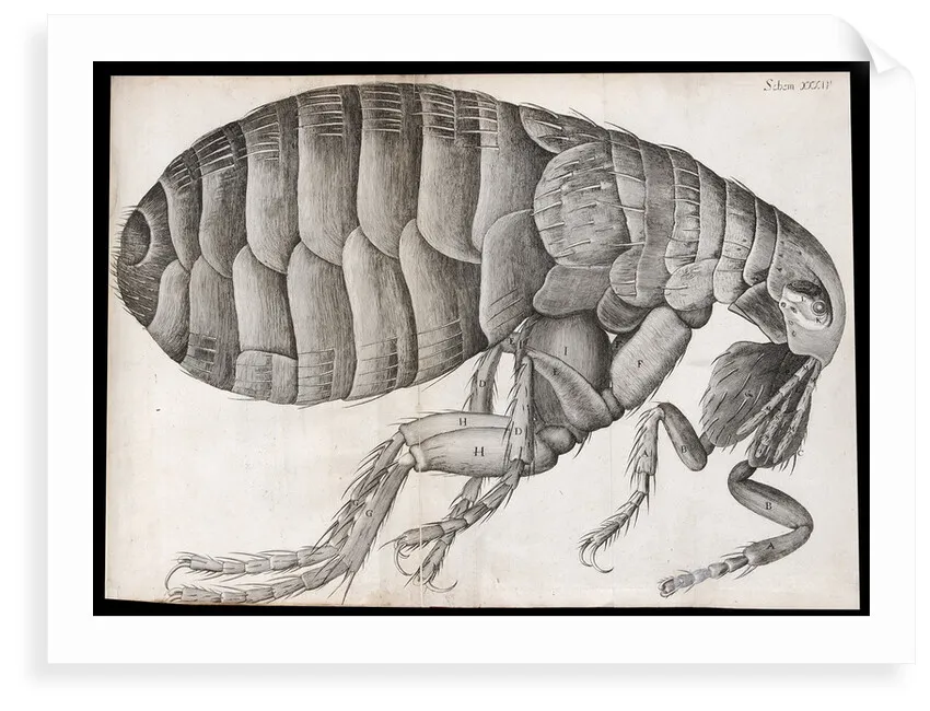
The book contained remarkably detailed illustrations of microscopic structures, many of which became iconic images in the history of science. One of the most famous examples is Hooke’s drawing of a flea, which appeared enormous, alien, and almost terrifying when magnified. Readers of the 17th century were astonished to discover that ordinary tiny creatures possessed such intricate anatomical complexity.
Another historically important observation came from Hooke’s examination of cork tissue. While studying thin slices of cork, he noticed countless tiny box-like compartments. He described these structures as “cells” because they reminded him of small rooms occupied by monks in monasteries.
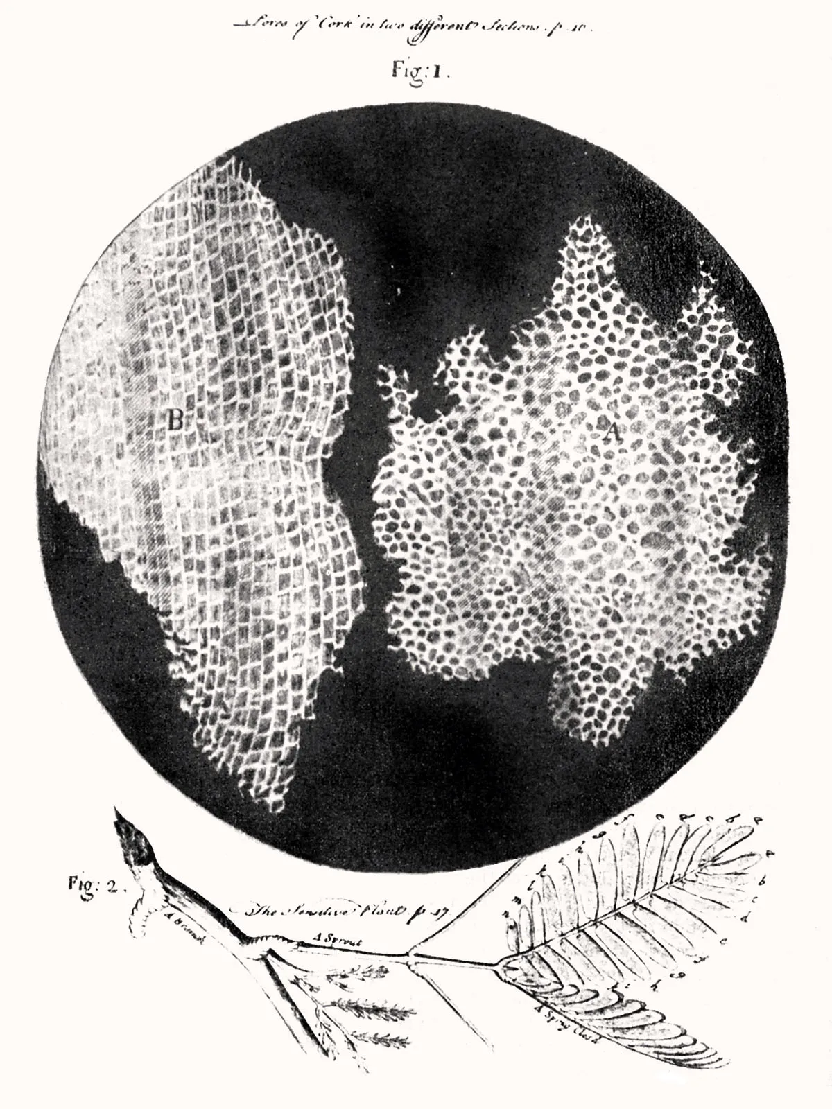
This was the first recorded scientific use of the word “cell” in biology. Although Hooke did not fully understand the biological importance of cells at the time, the concept later became one of the foundations of modern biology. In many ways, Micrographia was more than a scientific publication. It was also an artistic and philosophical work. Hooke’s illustrations transformed invisible microscopic structures into visual experiences that ordinary people could finally understand and imagine.
The book had a massive impact on European science and culture. Even people who were not scientists became fascinated by the hidden worlds revealed through magnification. Microscopy suddenly became exciting, mysterious, and intellectually fashionable. This historical moment still connects directly to modern digital microscopy today.
When modern microscope cameras display microscopic organisms on giant monitors, when AI systems analyze cellular structures, or when high-resolution CMOS sensors reveal invisible detail, they continue the same basic mission that Hooke began centuries ago:
extending human vision beyond the natural limits of the eye. The difference is that Hooke relied on hand drawings and primitive optics, while modern microscopy combines advanced lenses, digital imaging, computation, and artificial intelligence. Yet the sense of wonder remains remarkably similar. Even today, looking through a microscope — whether optical or digital — still carries something of the same feeling that readers experienced when opening Micrographia for the first time nearly 360 years ago.
Film Microscopy and the Birth of Scientific Imaging
Before digital imaging, microscope photography depended on film. Photomicrography became increasingly sophisticated during the 20th century, especially in medicine and biological research. Researchers used specialized film cameras mounted on microscopes to document:
- cells,
- bacteria,
- tissue samples,
- crystals,
- and microscopic structures.
Film-based microscopy produced remarkable images, but the process had limitations.
Users needed:
- careful exposure control,
- chemical film development,
- darkroom processing,
- and precise lighting techniques.
Results were often delayed by hours or days. Capturing moving microorganisms or dynamic biological processes was especially challenging because film systems were not well suited for rapid live imaging. Still, film microscopy laid the foundation for modern scientific imaging. Many iconic scientific discoveries were documented using film-based microscope photography.
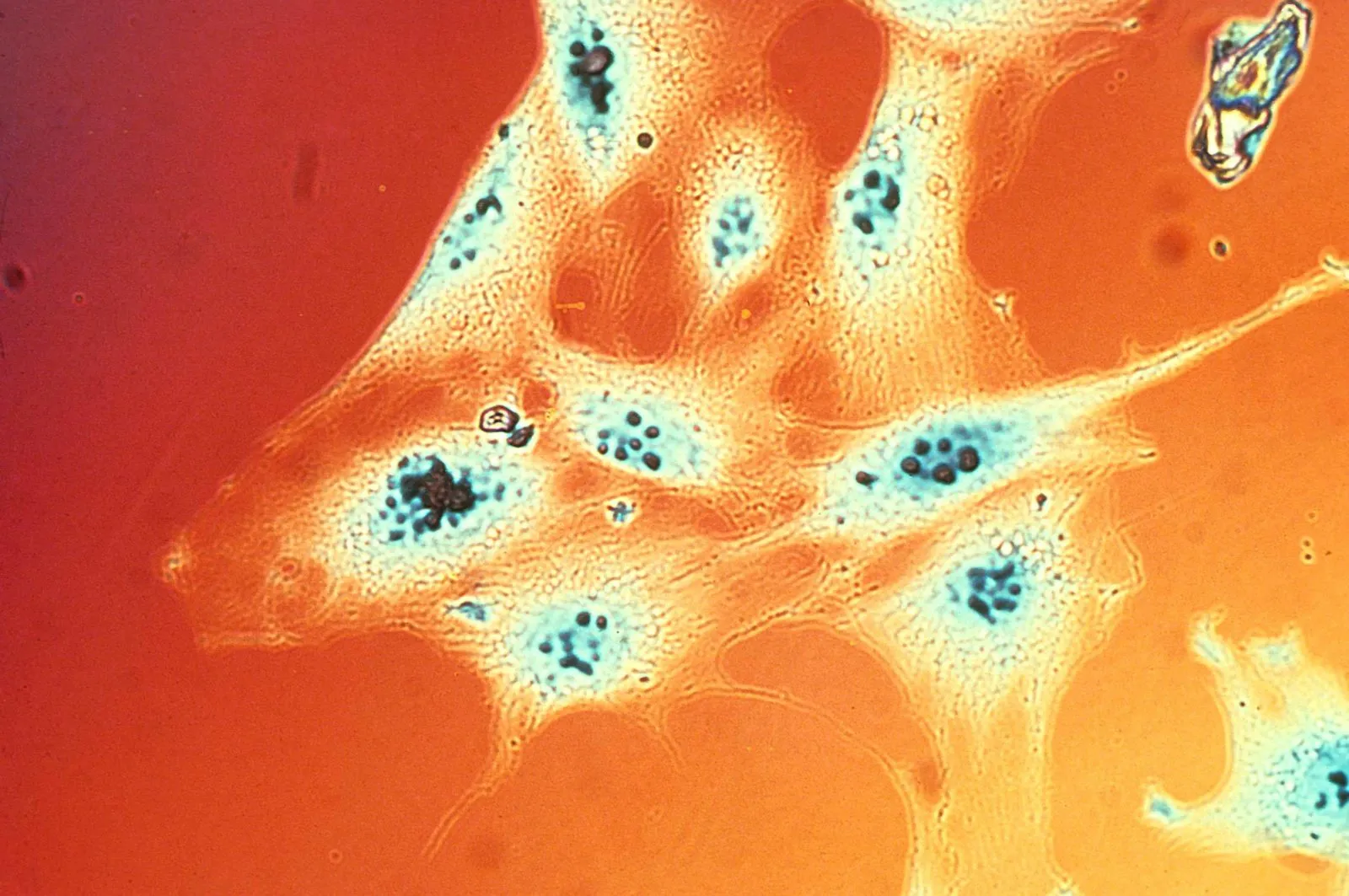
“Even iconic photomicrographs such as the 1976 Nikon Small World image Colony of Mouse Fibroblasts were created using traditional film photomicrography decades before digital microscope cameras became common.”
The Digital Revolution in Microscopy
The arrival of digital image sensors transformed microscopy completely.
Instead of recording light chemically onto film, digital sensors converted light directly into electronic signals. Images could now appear instantly on screens without chemical development. This changed microscopy in several major ways:
- immediate image review,
- live video observation,
- image storage,
- computer analysis,
- remote collaboration,
- and automated measurement.
Microscopy stopped being purely optical and became computational. The microscope was no longer just an instrument for viewing. It became a data-generation system.
CCD vs CMOS: The Sensor Evolution
Early digital microscope cameras often used CCD sensors. CCD stands for Charge-Coupled Device. For many years, CCD sensors were preferred in scientific imaging because they provided:
- excellent image quality,
- low electronic noise,
- high sensitivity,
- and good color performance.
However, CCD cameras also had disadvantages:
- high manufacturing cost,
- slower readout speeds,
- greater power consumption,
- and larger physical size.
Over time, CMOS sensor technology improved dramatically. CMOS stands for Complementary Metal-Oxide Semiconductor. Initially, CMOS sensors were considered inferior for scientific imaging. But modern CMOS technology evolved rapidly and eventually surpassed CCD systems in many applications. Today, CMOS sensors dominate digital microscopy because they offer:
- high resolution,
- fast frame rates,
- lower power consumption,
- lower cost,
- and compact design.
Modern scientific CMOS cameras can produce extraordinary image quality while operating at high speeds suitable for live observation and recording.
Why Resolution Is Not Everything
One of the biggest misconceptions in digital microscopy is the obsession with megapixels. Manufacturers often advertise cameras primarily by resolution:
- 5MP,
- 10MP,
- 20MP,
- or even higher.
Higher resolution certainly matters in some applications, but megapixels alone do not determine image quality. Several other factors are equally important:
- sensor size,
- pixel size,
- dynamic range,
- noise control,
- color accuracy,
- frame rate,
- optical quality,
- and software processing.
A poorly designed 20MP microscope camera may produce worse results than a well-designed lower-resolution system. Microscope imaging is especially demanding because microscopic subjects often contain:
- low-contrast detail,
- transparent structures,
- reflective surfaces,
- and difficult lighting conditions.
Good sensor engineering matters enormously.
Sensor Size and Why It Matters
Sensor size is one of the most overlooked specifications in microscope cameras. Larger sensors can:
- collect more light,
- improve dynamic range,
- reduce noise,
- and produce more natural imaging.
In microscopy, sensor size also affects the field of view. Small sensors may crop the microscope image significantly, making the captured image appear more “zoomed in” than the actual eyepiece view. Larger sensors preserve more of the optical field. This becomes especially important in:
- pathology,
- biological imaging,
- semiconductor inspection,
- and educational demonstrations.
Live Imaging and Real-Time Observation
One of the most transformative aspects of digital microscopy is live viewing. Instead of forcing users to observe through eyepieces individually, microscope cameras can display live images on monitors. This has enormous advantages.
- In education, an entire classroom can observe the same specimen simultaneously.
- In laboratories, multiple researchers can discuss observations together in real time.
- In industry, inspectors can examine components comfortably without prolonged eyepiece use.
- In medicine, specialists can collaborate remotely using live microscopic imaging.
The microscope becomes a shared visual platform rather than an isolated personal instrument.
Digital Measurement and Scientific Analysis
Modern microscope software can perform tasks impossible in traditional optical microscopy.
Digital systems now allow:
- distance measurement,
- area calculation,
- particle counting,
- edge detection,
- contrast enhancement,
- image stitching,
- and automated analysis.
This transformed microscopy from passive observation into quantitative analysis.
For example:
- metallurgists can measure grain structures,
- biologists can count cells automatically,
- semiconductor inspectors can analyze defects,
- and quality-control laboratories can document measurements precisely.
Software integration became as important as optics themselves.
Image Stacking and Extended Depth of Field
Microscopy faces a fundamental optical problem:
high magnification reduces depth of field. At high magnifications, only thin layers of the specimen remain in focus at one time. Digital imaging introduced a powerful solution: focus stacking.
In focus stacking, multiple images are captured at different focus positions and combined computationally into a single image with much greater apparent depth of field. This technique became extremely important in:
- insect photography,
- electronics inspection,
- gemology,
- and scientific illustration.
Modern software can automatically align and merge stacked images with remarkable precision. The result often appears far sharper and more detailed than what the human eye sees directly through the microscope.
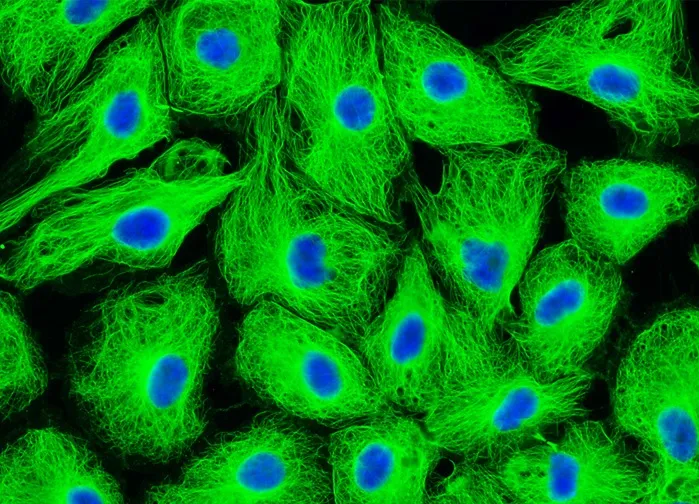
Fluorescence Microscopy and Digital Sensors
One of the most important applications of digital microscopy is fluorescence imaging. In fluorescence microscopy, specimens are tagged with fluorescent dyes that emit light when excited by specific wavelengths.
This allows researchers to visualize:
- proteins,
- DNA,
- cellular structures,
- bacteria,
- and biological activity.
Digital cameras are essential in fluorescence microscopy because fluorescence signals are often extremely faint. Sensitive scientific cameras can detect subtle light emissions invisible to the human eye. Modern biomedical research would be almost impossible without advanced digital microscope imaging systems.
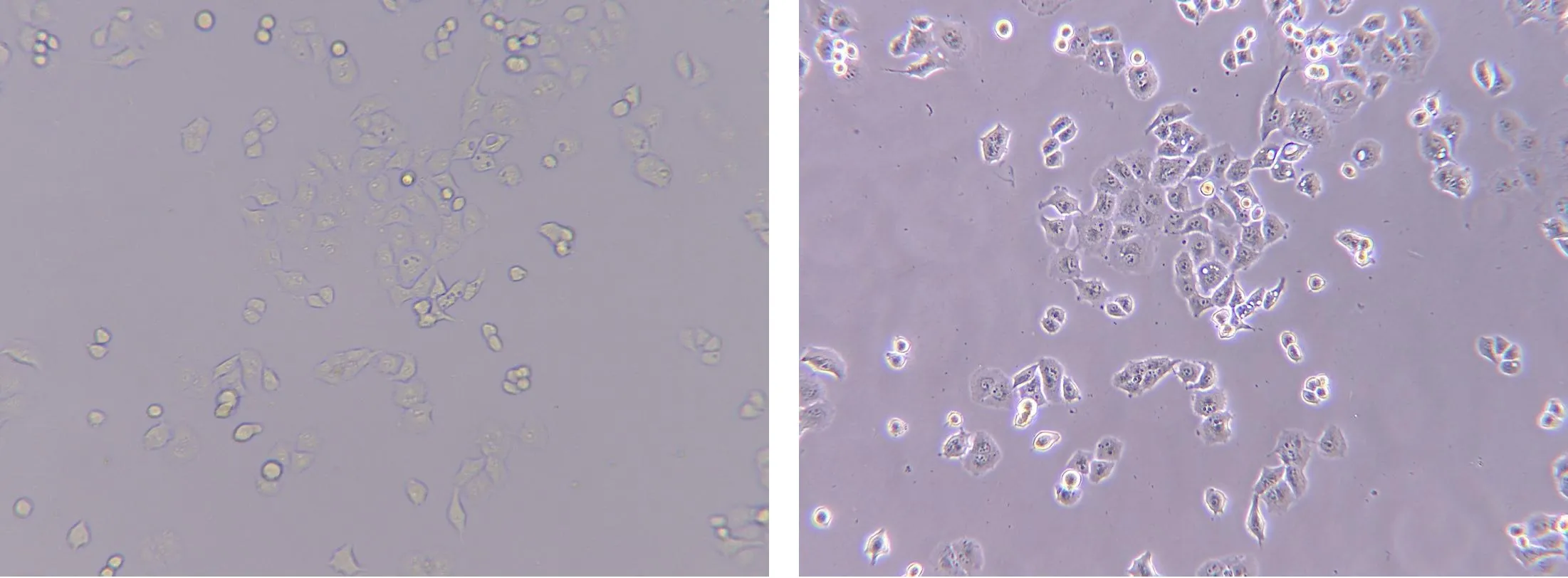
Digital Microscopy in Medicine
Medical microscopy changed dramatically after digital imaging became widespread. Pathology laboratories increasingly use digital slide scanning systems capable of converting entire microscope slides into ultra-high-resolution digital images. These systems allow:
- remote diagnosis,
- AI-assisted analysis,
- educational sharing,
- and long-term archiving.
A pathologist no longer necessarily needs physical access to the microscope itself. Digital pathology is becoming one of the most important fields in modern medicine. Artificial intelligence systems are now being trained to identify:
- cancer cells,
- blood abnormalities,
- tissue patterns,
- and infectious organisms.
The microscope camera became not just a recording tool, but part of an intelligent diagnostic system.
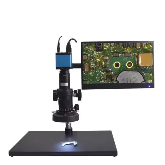
Industrial Microscopy and Electronics Inspection
Digital microscopy also transformed industry. Modern manufacturing increasingly depends on microscopic inspection. Industries using digital microscopy include:
- semiconductor manufacturing,
- electronics assembly,
- aerospace engineering,
- metallurgy,
- automotive production,
- and materials science.
PCB inspection is one of the most common examples. Digital microscope cameras allow technicians to inspect:
- solder joints,
- micro-components,
- circuit traces,
- connector pins,
- and manufacturing defects.
Live monitor viewing improves comfort dramatically during prolonged inspection work. Digital recording also creates documentation for quality control and traceability.
Education and Accessibility
Digital microscopy made microscopy more accessible than ever before. Students no longer need to crowd around a single eyepiece. Schools can project live microscopic images onto screens or interactive displays. Online education also became far easier.
Teachers can now:
- stream microscope demonstrations,
- record laboratory exercises,
- and share digital specimen libraries globally.
Even amateur microscopists benefit enormously from digital cameras because observations can easily be:
- photographed,
- archived,
- discussed online,
- and shared with scientific communities.
Microscopy became more collaborative and social.
The Rise of USB Microscope Cameras
USB microscope cameras played a major role in popularizing digital microscopy.
Earlier scientific camera systems were often expensive and required specialized hardware.
USB cameras simplified everything.
Users could connect microscope cameras directly to ordinary computers and immediately begin capturing images.
This dramatically lowered the barrier to entry.
Modern USB microscope cameras now offer:
- high resolution,
- live video,
- measurement tools,
- image capture,
- and software integration
at relatively affordable prices.
Why Dedicated Microscope Cameras Still Matter
Some people wonder why dedicated microscope cameras are necessary when smartphones have excellent cameras.
The answer is that microscope imaging has unique requirements.
Dedicated microscope cameras are designed specifically for:
- microscope optics,
- scientific color accuracy,
- low-light imaging,
- sensor compatibility,
- and prolonged live operation.
Smartphones can work surprisingly well in some situations, but dedicated microscope cameras remain superior for serious microscopy because they provide:
- stable mounting,
- optimized sensors,
- scientific software,
- precise scaling,
- and better integration with microscope systems.
Artificial Intelligence and the Future of Microscopy
The future of digital microscopy increasingly involves artificial intelligence.
AI systems are already being trained to:
- identify microorganisms,
- classify cells,
- detect cancer,
- analyze tissue samples,
- count particles,
- and identify manufacturing defects.
In some applications, AI can already detect patterns invisible to inexperienced human observers. This does not necessarily mean microscopes will replace scientists or doctors. Instead, AI will likely become an analytical assistant that improves speed, consistency, and diagnostic accuracy.
The combination of:
- digital sensors,
- high-speed computing,
- cloud systems,
- and machine learning
is rapidly transforming microscopy into a semi-automated analytical science.
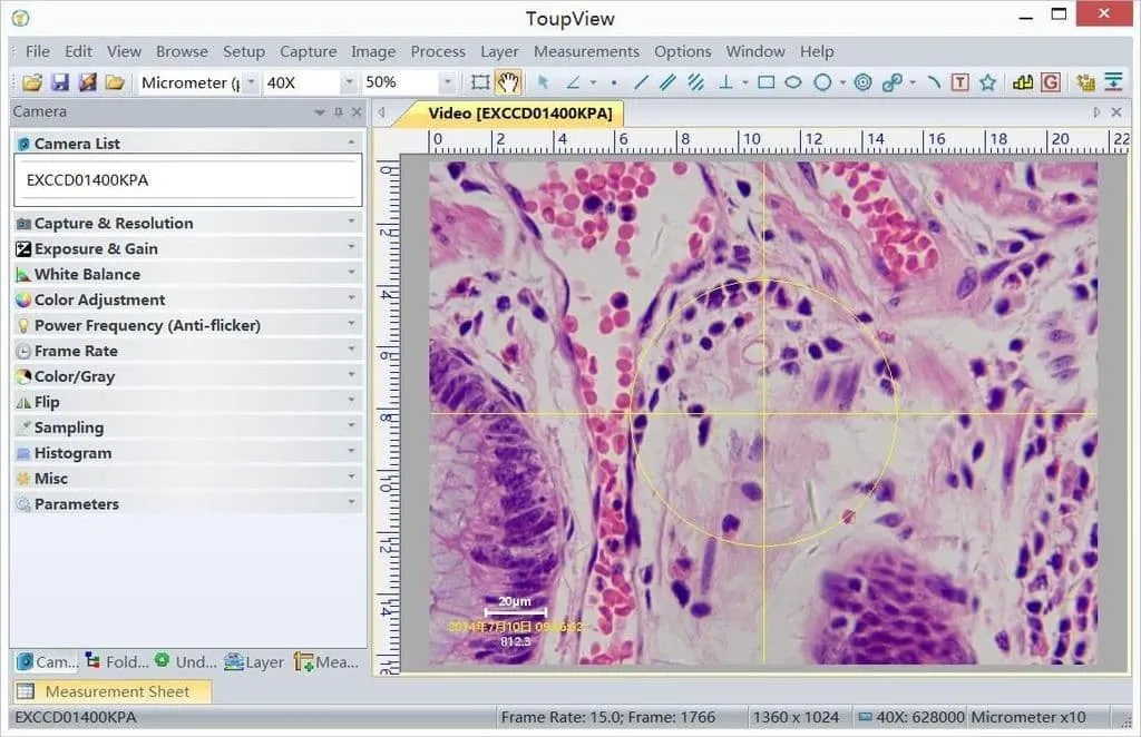
The Importance of Software
Modern microscope cameras are no longer just hardware devices. Software quality is now critically important. Good microscopy software allows users to:
- capture images,
- record video,
- calibrate measurements,
- adjust exposure,
- control white balance,
- create image stacks,
- annotate observations,
- and manage scientific data.
Poor software can make even excellent hardware frustrating to use. This is why serious microscope camera manufacturers invest heavily in software ecosystems alongside sensor technology.
ToupTek and Modern Microscope Camera Manufacturers
Today, several manufacturers specialize in digital microscopy imaging systems.
Among them, ToupTek became increasingly well known for producing microscope cameras that balance affordability, functionality, and modern imaging performance.
ToupTek cameras are widely used in:
- education,
- laboratories,
- industrial inspection,
- biological microscopy,
- and research environments.
One of the company’s strengths is the wide variety of camera models available for different applications, including:
- USB microscope cameras,
- HDMI microscope cameras,
- high-resolution imaging systems,
- and scientific CMOS cameras.
The ToupTek E3ISPM20000KPA Microscopy Camera is one example of how modern microscope cameras evolved beyond simple imaging accessories into advanced digital observation systems capable of high-resolution live imaging and scientific documentation.
Other important manufacturers in the digital microscopy world include:
- Olympus Corporation,
- Leica Microsystems,
- Nikon,
- Carl Zeiss,
- AmScope,
- and Motic.
Each company approaches digital microscopy differently depending on whether the focus is:
- education,
- industrial inspection,
- pathology,
- fluorescence imaging,
- or advanced scientific research.
Conclusion: Microscopy Became Digital, Collaborative, and Intelligent
The history of microscopy is ultimately the history of extending human vision.
From simple lenses to electron microscopes, every improvement allowed humanity to observe previously invisible worlds.
Digital microscopy represents one of the most important steps in that evolution because it transformed microscopy from a solitary optical experience into a digital, shareable, measurable, and increasingly intelligent system.
Microscope cameras changed not only how scientists record images, but also how knowledge itself is created, analyzed, shared, and preserved.
Today, a modern microscope camera can:
- stream live biological activity,
- analyze microscopic structures,
- archive scientific evidence,
- support AI-assisted diagnostics,
- and connect researchers globally in real time.
The microscope is no longer simply an instrument for seeing.
It became an instrument for data, communication, and computational discovery.
And as sensor technology, artificial intelligence, and imaging software continue evolving, digital microscopy will almost certainly become even more powerful — revealing details, patterns, and relationships that earlier generations of scientists could barely imagine.

No Comments