


Key Features
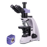
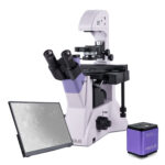
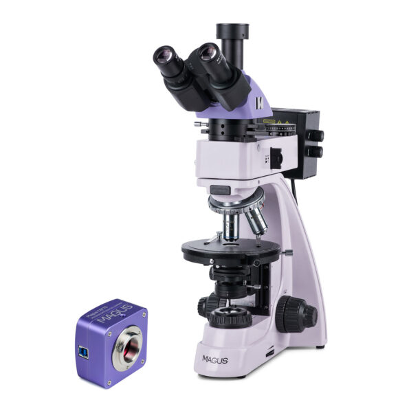
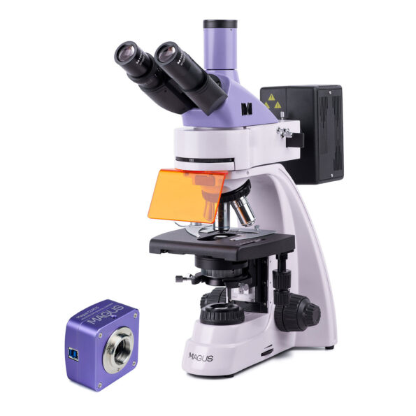
MAGUS Bio VD350 Biological Inverted Digital Microscope
Key Features of the Microscope
- Sample Viewing: Designed for viewing samples in laboratory ware up to 55mm high and with a bottom thickness of up to 1.2mm. Tilt the illuminator stand to accommodate dishes up to 165mm high.
- Microscopy Techniques: Supports brightfield and phase-contrast techniques, featuring a phase-contrast condenser with a field diaphragm.
- Stage and Movement: Includes a stage with 4 dish holders of varying sizes, and a mechanical attachment for moving the sample along two horizontal axes.
- Rotatable Head: The microscope head rotates 180° for versatile viewing angles.
- Camera and Monitor Mounting: Offers two mounting options for the camera and monitor: in the vertical tube or on the microscope body.
- Illumination: Uses a 30W halogen lamp as a transmitted light source, powered by an AC power supply.
- Accessories: Compatible with a wide range of optional accessories to enhance functionality.
Key Features of the Camera
- High Sensitivity Sensor: Equipped with a SONY Exmor backlit color CMOS sensor for high light sensitivity.
- Versatile Operation: Functions in darkfield and brightfield with 4x, 10x, and 20x objectives.
- Fast Data Transfer: Connects to a computer via a USB3.0 interface, offering a data transfer speed of 5Gbps.
- High-Resolution Output: Outputs images to a monitor in ultra-high resolution 4K (3840x2160px) at 45fps or Full HD (1920x1080px) at 70fps.
- Included Software: Comes with drivers and image processing software for easy setup and use.
- Flexible Installation: Can be installed into the trinocular tube or the eyepiece tube of a microscope.
Description
MAGUS Bio VD350 Biological Inverted Digital Microscope
The MAGUS Bio VD350 is a high-performance biological inverted microscope, perfect for examining both stained and unstained samples in laboratory containers such as Petri dishes, flasks, and plates. It accommodates dishes up to 55mm in height, and with the illuminator stand tilted, it can handle dishes up to 165mm high. The microscope supports containers with a bottom thickness of up to 1.2mm. Utilizing both brightfield and phase contrast microscopy techniques in transmitted light, this microscope is ideal for routine laboratory research, teaching, and various scientific applications. It features a trinocular tube and side port for mounting digital cameras and monitors, enhancing its versatility.
Digital Camera
The MAGUS CDF30 digital camera features a SONY Exmor 8.3MP sensor with back-illumination technology, ensuring high light sensitivity and producing bright, clear images even in low-light conditions. This camera is suitable for both darkfield and brightfield microscopy techniques using 4x, 10x, and 20x objectives, and is also compatible with stereomicroscopes. It connects to a computer via a high-speed USB3.0 port, which supports data transfer rates up to 5Gbps, enabling seamless image display on the screen.
The camera can be installed in the trinocular tube or eyepiece tube of the microscope. For the best field of view without optical defects, use an adapter with a magnification between 0.65x and 0.9x. Additional adapters are needed for eyepiece tube installation.
The camera delivers images at a maximum resolution of 3840x2160px with a frame rate of 45fps. At a lower resolution of 1920x1080px, the frame rate increases to 70fps, ensuring smooth transitions without delays. This makes it suitable for studying and presenting moving objects. Photos and videos can be recorded and saved in various formats.
Optics
The MAGUS Bio VD350 microscope's trinocular head can be rotated 180° to adjust the height of the exit pupil. The vertical tube is used to mount a monitor, while the side port on the microscope body is used for mounting a digital camera. Beam splitting on the body is adjustable between 100/0 and 0/100, with options for 50/50 on the trinocular tube when open, or 100/0 when closed.
The microscope includes four infinity plan achromatic objectives, one of which is a phase objective. The revolving nosepiece accommodates four objectives, leaving one slot free for an additional objective.
Illumination
The phase-contrast condenser allows for easy switching between microscopy techniques. It has four working positions: a free aperture for brightfield and 10x, 20x, and 40x apertures for phase contrast with the appropriate magnification objective. The transmitted light source is a 30W halogen bulb with adjustable brightness, powered by an AC supply. The illumination system supports Köhler illumination, enhancing image quality.
Stage and Focusing Mechanism
The microscope comes with four dish holders for the stage, which can be moved along the X and Y axes using a mechanical attachment, while the stage remains stationary. The holders are designed to accommodate various sizes of labware. The focusing mechanism operates smoothly, with a coarse focusing lock knob and tension adjustment. The fine focusing scale value is 2μm, and the focusing knobs are located at the base for ergonomic operation.
Accessories
A wide range of optional accessories, including additional eyepieces, phase objectives, digital cameras, and calibration slides, are available to maximize the capabilities of the MAGUS Bio VD350 microscope.
The kit contains:
- MAGUS CDF30 Digital Сamera (digital camera, USB cable, installation CD with drivers and software, user manual and warranty card)
- Stand with the built-in power supply, transmitted light source, focusing mechanism, stage, condenser mount, revolving nosepiece, and trinocular tube
- Phase-contrast condenser
- Trinocular head
- Infinity plan achromatic objective: PHP2 10x/0.25 phase WD 4.27mm
- Infinity plan achromatic objective: PL 10x/0.25 WD 4.27mm
- Infinity plan achromatic objective: PL 20x/0.40 WD 8.0mm
- Infinity plan achromatic objective: PL 40x/0.60 WD 3.5mm
- Eyepiece 10x/22mm with long eye relief (2 pcs.)
- Eyecup (2 pcs.)
- Centering telescope
- Round stage plate
- Mechanical attachment for moving the specimen
- Dish holder (4 pcs.)
- C-mount camera adapter
- Color filter
- AC power cord
- Dust cover
- User manual and warranty card
Available on demand:
- 10x/22mm eyepiece with a scale
- 12.5x/14mm eyepiece (2 pcs.)
- 15x/15mm eyepiece (2 pcs.)
- 20x/12mm eyepiece (2 pcs.)
- 25x/9mm eyepiece (2 pcs.)
- Infinity plan achromatic objective: PL 4x/0.10 WD 21mm
- Infinity plan achromatic objective: PHP2 20x/0.40 phase WD 8.0mm
- Infinity plan achromatic objective: PHP2 40x/0.60 phase WD 3.5mm
- Calibration slide
- LCD Monitor
specifications:
| Product ID | 83014 |
| Brand | MAGUS |
| Warranty | 5 years |
| EAN | 5905555018126 |
| Package size (LxWxH) | 19.3x13.4x28.7 cm |
| Shipping Weight | 44.3 kg |
| Microscope specifications | |
| Type | biological, light/optical, digital |
| Head | trinocular |
| Nozzle | Siedentopf, rotatable 180° |
| Head inclination angle | 45 ° |
| Magnification, x | 100–400 basic configuration (*optional: 40–500/600/800/1000) |
| Eyepiece tube diameter, in | 1.2 |
| Eyepieces | 10х/22mm, eye relief: 10mm (*optional: 10x/22mm with scale, 12.5x/14; 15x/15; 20x/12; 25x/9) |
| Objectives | infinity plan achromatic: PL 10x/0.25, PL 20x/0.40, PL 40x/0.60, PHP2 10x/0.25 phase; parfocal distance 45mm (*optional: PL 4x/0.10, PHP2 20x/0.40 phase, PHP2 40x/0.60 phase) |
| Revolving nosepiece | for 5 objectives |
| Working distance, mm | 4,27 (10x); 8,0 (20х); 3,5 (40x) |
| Interpupillary distance, in | 1.9 — 3 |
| Stage, mm | 227x208 |
| Stage moving range, mm | 77/114 |
| Stage features | fixed, with glass plate Ø118mm and mechanical attachment; dish holders: 86x129.5, Ø90mm; 34x77.5mm, Ø68.5mm; 57x82mm, Ø60mm; 29x77.5mm, Ø35mm |
| Condenser | NA 0.6, working distance: 55mm; phase contrast turret |
| Diaphragm | adjustable aperture diaphragm, adjustable iris field diaphragm |
| Focus | coaxial, coarse (with coarse focusing tension adjustment and a lock knob), and fine (0.002mm) |
| Illumination | halogen |
| Brightness adjustment | ✓ |
| Power supply | 220±22V, 50Hz, AC network |
| Light source type | 12V/30W |
| Light filters | yes |
| Operating temperature range, °F | 41...+95 |
| Special features | phase contrast condenser (turret) with a free slot and phase annuli plates for 10x, 20x and 40x objectives; centering auxiliary microscope |
| User level | experienced users, professionals |
| Assembly and installation difficulty level | complicated |
| Application | laboratory/medical |
| Illumination location | lower |
| Research method | bright field, phase-contrast microscopy |
| Pouch/case/bag in set | dust cover |
| Camera specifications | |
| Sensor | SONY Exmor CMOS |
| Color/monochrome | color |
| Megapixels | 8.3 |
| Maximum resolution, pix | 3840x2160 |
| Sensor size | 1/1.2'' (11.14x6.26mm) |
| Pixel size, μm | 2.9x2.9 |
| Light sensitivity | 5970mV/lux/s |
| Signal/noise ratio | 0.13mV at 1/30s |
| Exposure time | 0.02ms–15s |
| Video recording | ✓ |
| Video recording | yes |
| Frame rate, fps at resolution | 45@3840x2160, 70@1920x1080 |
| Place of installation | trinocular tube, eyepiece tube instead of an eyepiece |
| Image format | *.jpg, *.bmp, *.png, *.tif |
| Video format | output: *.wmv, *.avi, *.h264 (Windows 8 and later), *h265 (Windows 10 and later) |
| Spectral range, nm | 380–650 (built-in IR filter) |
| Shutter type | ERS (electronic rolling shutter) |
| White balance | manual, automatic |
| Exposure control | manual, automatic |
| Software | MAGUS View |
| Output | USB 3.0, 5Gb/s |
| System requirements | Windows 8/10/11 (32bit and 64bit), Mac OS X, Linux, up to 2.8GHz Intel Core 2 or higher, minimum 2GB RAM, USB3.0 port, CD-ROM, 17" or larger display |
| Mount type | C-mount |
| Body | solid aluminum |
| Camera power supply | DC, 5V, from computer USB port; a 12V, 3A adapter for Peltier element |
| Camera operating temperature range, °F | -10...+50 |
| Operating humidity range, % | 30 — 80 |
Reviews
Recommended


0.0 Average Rating Rated (0 Reviews)