The Ultimate Guide to Different Types of Microscope
-
 BiGGie
- NightSky
- No Comments

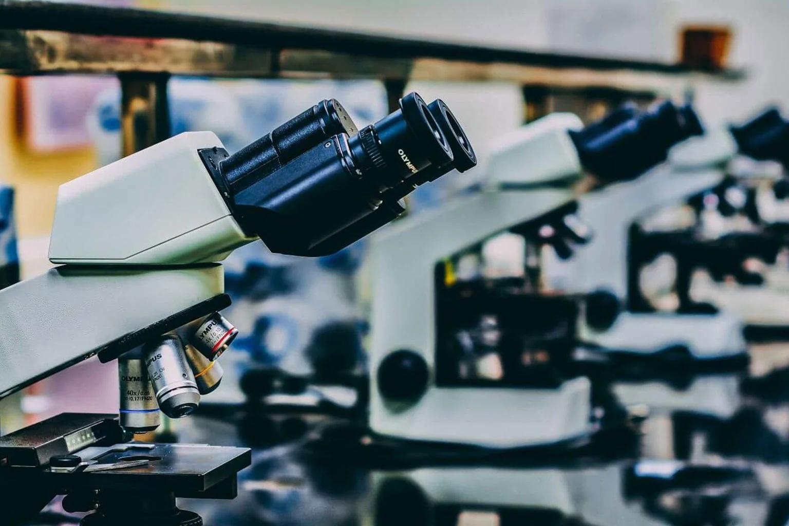
Table of Contents
Understanding the Types of Microscopes and Their Functions
Our world contains countless invisible wonders, from the tiniest bacteria to the intricate structures of cells. These hidden realms become accessible through various types of microscopes, powerful tools that reveal what our eyes cannot perceive.
Modern science recognizes several microscope classifications, each containing specialized microscope varieties tailored for specific applications. Whether examining living organisms or analyzing material structures, these instruments are precisely engineered to meet different scientific needs.
For example:
- Compound microscopes are best for viewing thin, transparent samples like cells on a slide.
- Stereo microscopes provide a 3D view of larger, solid objects such as insects, plant parts, and circuit board components.
- Electron microscopes use beams of electrons instead of light to achieve much higher magnification, allowing researchers to see viruses or nanostructures.
A Breakdown of the Main Types of Microscopes
Microscopes play a crucial role in scientific discovery, enabling researchers to observe structures too small for the naked eye. They can be classified into several types based on their working principles and applications.
1. Optical Microscopes (Light Microscopes)
These are the most commonly used microscopes in laboratories and educational settings.
- Compound Microscope
- Uses multiple lenses to magnify specimens (objective & eyepiece lens).
- Ideal for viewing thin sections of biological samples.
- Commonly used in biology, medicine, and materials science.
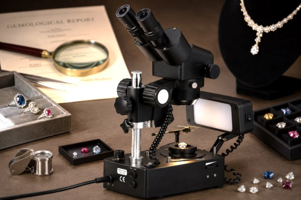
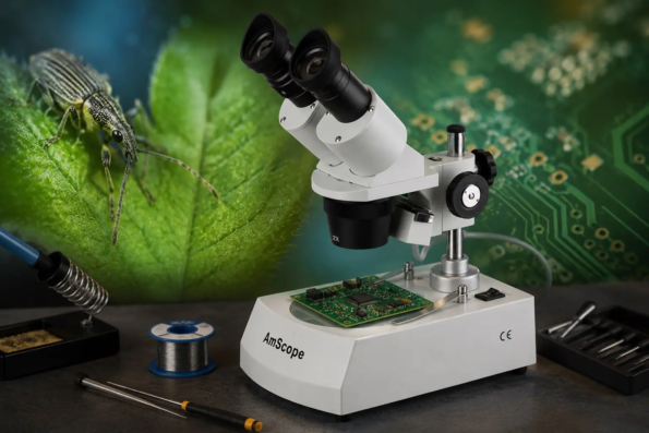
- Stereo Microscope (Dissecting Microscope)
- Provides a 3D view of specimens at lower magnifications.
- Used in dissections, circuit board inspections, and material studies.
- Does not require sample preparation like slides.
2. Electron Microscopes
Designed for extremely high-resolution imaging, electron microscopes use electron beams instead of light.
- Transmission Electron Microscope (TEM)
- Produces high-resolution 2D images by transmitting electrons through the sample.
- Used in nanotechnology, virology, and material science.
- Requires ultra-thin sample sections.
- Scanning Electron Microscope (SEM)
- Produces detailed 3D surface images by scanning a focused electron beam over a sample.
- Essential for industrial analysis, metallurgy, and biological research.
3. Scanning Probe Microscopes
These microscopes use a physical probe to scan surfaces at the atomic level.
- Atomic Force Microscope (AFM)
- Measures surface interactions at an atomic scale.
- Used in nanotechnology, semiconductor research, and molecular biology.
- Scanning Tunneling Microscope (STM)
- Used for visualizing individual atoms.
- Critical for research in quantum mechanics and material engineering.
Choosing the Right Microscope
The choice of microscope depends on magnification needs, sample type, and application:
- Biological studies: Compound microscopes or TEM.
- Material science & engineering: SEM or STM.
- Nanotechnology: AFM or TEM.
- Educational use: Optical microscopes.
Types of Optical Microscopes: From Brightfield to Fluorescence
Optical microscopes utilize visible light and lenses to magnify objects, making them essential in laboratories, research, and diagnostics. Different types cater to specific applications based on illumination techniques and contrast methods.
1. Brightfield Microscopy (Standard Optical Microscope)
This is the most common and basic type of optical microscope.
- How it Works: Light passes through the sample, creating contrast between the specimen and the background.
- Best for: Stained biological samples like cells, tissues, and bacteria.
- Limitations: Struggles with transparent, unstained specimens due to low contrast.
2. Dark Field Microscopy
Enhances contrast by blocking direct light, illuminating only scattered light from the specimen.
- How it Works: The condenser prevents direct light from entering the objective, making the specimen appear bright against a dark background.
- Best for: Observing unstained, live organisms and fine structures like bacteria and blood cells.
- Advantage: Produces high-contrast images without needing dyes or stains.
Alt: Phase-Contrast Microscopy: Principles and Applications
Image credit: iabdm.org
3. Phase-Contrast Microscopy
Ideal for viewing live cells and transparent specimens without staining.
- How it Works: Uses a special phase plate to enhance differences in light wave interference, making subtle cellular structures visible.
- Best for: Examining internal cell structures, organelles, and living microorganisms.
- Advantage: Allows observation without chemical staining, preserving biological integrity.
4. Differential Interference Contrast (DIC) Microscopy (Nomarski Microscopy)
Creates 3D-like images with enhanced depth and contrast.
- How it Works: Uses polarized light to highlight variations in specimen thickness and density.
- Best for: High-resolution imaging of cells, embryos, and intricate biological details.
- Advantage: Provides sharp, relief-like images, making structures stand out.
5. Fluorescence Microscopy
Revolutionary for biological and medical research, allowing high-specificity imaging with fluorescent markers.
- How it Works: Specific wavelengths of light excites fluorescent molecules in the sample, causing them to emit bright, colored light against a dark background.
- Best for: Studying proteins, DNA, viruses, and molecular interactions.
- Advantage: Enables real-time tracking of live biological processes using fluorescent dyes.
Types of Electron Microscopes and their Scientific Applications
Electron microscopes, one of the most potent kinds of microscopes, use beams of electrons instead of light to achieve much higher magnifications and resolutions.
These instruments are essential for nanotechnology, biology, and materials science. Within microscope classifications, there are two main varieties of electron microscopes, each designed for specific applications.
1. Transmission Electron Microscope (TEM)
TEM is the most powerful tool for observing internal structures at the nanoscale.
- How it Works:
- Electrons pass through an ultra-thin specimen, interacting with its structures.
- A detector captures transmitted electrons to create high-resolution, 2D images.
- Key Applications:
- Nanotechnology – Identifies atomic structures of nanoparticles and quantum materials.
- Cell Biology & Virology – Studies viruses, bacteria, and organelle structures with sub-nanometer resolution.
- Material Science – Examines crystal structures, defects, and semiconductor materials.
- Advantages:
- Delivers ultra-high resolution down to the atomic level.
- Essential for analyzing internal fine details of cells and materials.
2. Scanning Electron Microscope (SEM)
SEM provides detailed 3D surface imaging, making it ideal for material analysis and biological research.
- How it Works:
- A focused electron beam scans the surface of the sample.
- Detectors collect reflected electrons, generating high-contrast, topographical images.
- Key Applications:
- Industrial Quality Control – Analyzes surface defects in metals, polymers, and electronics.
- Biological Research – Investigates cell surfaces, pollen grains, and insect morphology.
- Forensics & Archaeology – Examines artifacts, fingerprints, and forensic samples with precision.
- Advantages:
- Produces detailed surface images with depth perception.
- Useful for studying microstructures and textures.
Specialized Electron Microscopes
Beyond TEM and SEM, advanced variants enable even more specialized imaging:
- Scanning Transmission Electron Microscope (STEM)
- Hybrid technology combining SEM and TEM for high-resolution imaging with elemental analysis.
- Used in chemistry, material engineering, and drug development.
- Cryo-Electron Microscopy (Cryo-EM)
- Allows imaging of biological molecules in their natural, frozen state.
- Revolutionizing structural biology, helping map proteins and viruses.
Alt: Scanning Probe Microscopy: Principles & Applications
Image credit: mcf.gatech.edu
Exploring Scanning Probe Microscopes: A New Class of Microscopy
Scanning Probe Microscopes (SPMs) represent a revolutionary leap in microscopy, enabling researchers to explore surfaces at atomic and molecular scales.
Unlike traditional optical or electron microscopes, SPMs use physical probes to scan surfaces with exceptional precision, making them indispensable for nanotechnology, materials science, and molecular biology.
1. How Scanning Probe Microscopes Work
- Utilize an extremely fine probe that moves across the surface of a sample.
- The probe interacts with the surface through mechanical, electrical, or magnetic forces, generating precise topographical images.
- Provide nanoscale resolution, revealing structures down to individual atoms.
2. Main Types of Scanning Probe Microscopes
Atomic Force Microscope (AFM)
- Measures tiny forces between the probe and the sample surface.
- Generates high-resolution 3D images, displaying surface roughness and structural details.
- Used in materials science, semiconductor research, and biological studies.
- Advantage: Works in air, liquid, or vacuum, making it versatile for different environments.
Scanning Tunneling Microscope (STM)
- Explores electronic properties at the atomic level using quantum tunneling principles.
- Provides unparalleled resolution, capable of visualizing individual atoms.
- Used in quantum mechanics, surface science, and nanotechnology research.
- Advantage: Enables direct manipulation of single atoms, advancing molecular engineering.
Other Specialized SPM Variants
- Magnetic Force Microscopy (MFM): Maps magnetic domains crucial for data storage research.
- Electrochemical Scanning Probe Microscopy (EC-SPM): Investigates electrochemical reactions, vital for battery technology and corrosion studies.
3. Scientific Applications of Scanning Probe Microscopy
SPMs have transformed research across multiple disciplines, including:
- Nanotechnology & Materials Science: Analyzes atomic structures, aiding in the development of stronger, more efficient materials.
- Biological & Medical Research: Investigates protein structures, DNA interactions, and cellular membranes at molecular resolution.
- Semiconductor & Electronics Industry: Used for precise surface analysis in chip manufacturing and nanoelectronics.
- Environmental Science & Chemistry: Examines chemical compositions, corrosion mechanisms, and pollutant interactions at the nanoscale.
4. Why Scanning Probe Microscopes Are a Game-Changer
- Provide ultra-high resolution, surpassing even electron microscopy for surface analysis.
- Enable non-destructive imaging, preserving delicate biological and material samples.
- Offer multi-functional capabilities, combining topographic mapping with electrical, magnetic, and mechanical data collection.
Modern Types of Microscopes: Digital and AI-Powered Technologies
Microscope classifications have expanded significantly as microscopy advances beyond traditional optical and electron techniques.
Today's microscope varieties include cutting-edge microscopes that incorporate digital imaging, artificial intelligence (AI), and automation. These innovations are enhancing precision, efficiency, and accessibility in scientific research and industrial applications.
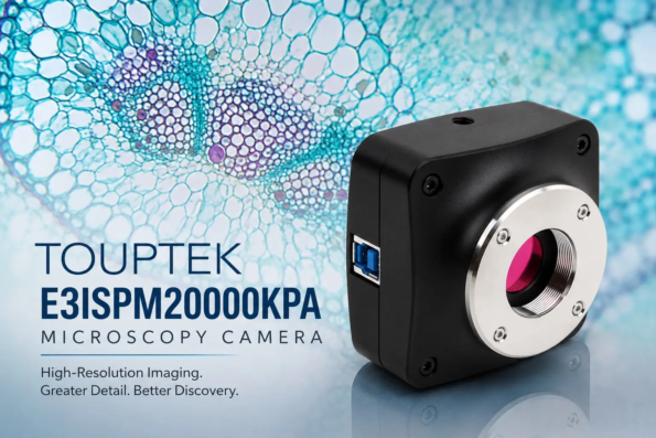
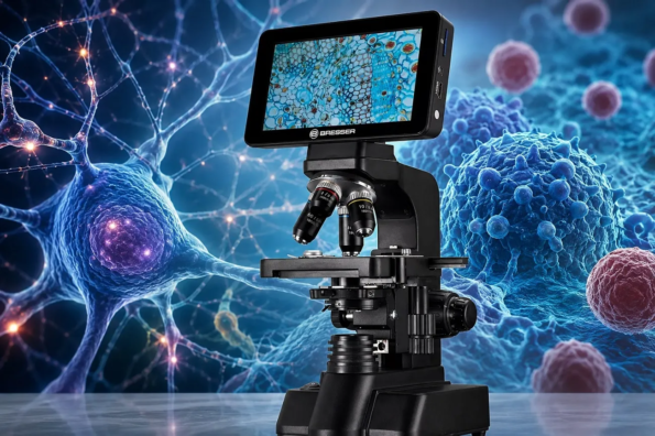
1. Digital Microscopes
Digital microscopes replace traditional eyepieces with high-resolution cameras and digital displays, offering enhanced visualization and analysis.
- Key Features:
- Live imaging & recording: Captures images in real-time for analysis and sharing.
- High-definition display: Eliminates optical distortion, providing clearer views.
- Software integration: Enables measurement, annotation, and automated image analysis.
- Types of Digital Microscopes:
- USB Digital Microscopes: Compact and portable, used in education, quality control, and electronics inspection.
- Wireless Digital Microscopes: Allows remote viewing via Wi-Fi or mobile devices, ideal for collaborative research and field studies.
- Automated Digital Microscopes: High-throughput systems for biomedical research, materials science, and industrial applications.
Alt: AI-Powered Microscopes: Smart Imaging for Advanced Research
Image credit: www.endava.com
2. AI-Powered Microscopes
The integration of artificial intelligence has transformed microscopy, enabling automated image analysis, anomaly detection, and predictive insights.
- How AI Enhances Microscopy:
- Pattern recognition: Identifies structures and abnormalities with precision beyond human analysis.
- Automated diagnostics: Speeds up research in medicine, pathology, and drug discovery.
- Real-time processing: Reduces the need for manual interpretation, improving efficiency.
- Applications of AI Microscopy:
- Medical Imaging & Pathology: AI assists in detecting cancer cells, tissue abnormalities, and microbial infections.
- Materials Science & Nanotechnology: AI analyzes surface defects, molecular interactions, and chemical compositions.
- Environmental Science: Tracks microbial ecosystems, pollution patterns, and climate-related biological changes.
3. Hybrid Smart Microscopes
The latest hybrid technologies combine digital imaging, AI, and robotics, offering enhanced capabilities.
- Examples of Smart Microscopy Systems:
- Deep-Learning Microscopes: AI-driven tools for advanced cell analysis, molecular research, and neuroscience.
- Automated 3D Microscopes: Capture three-dimensional cellular structures essential for tissue engineering and regenerative medicine.
- Cloud-Connected Microscopes: Remote microscopy for collaborative research and global accessibility.
Why Modern Microscopy is a Game-Changer
- Improved accuracy: Reduces human errors in medical diagnostics and industrial inspections.
- Faster processing: Enables high-throughput analysis, saving time in research and production.
- Global accessibility: Allows remote collaboration using cloud-based tools.
Comparison of All Types of Microscopes: Features, Cost, and Use Cases
Microscope classifications show that these essential tools come in many microscope varieties, each with unique features, costs, and applications across scientific disciplines.
Choosing the right type depends on resolution needs, sample properties, and budget constraints. Below is a structured comparison of major microscope types.
1. Optical Microscopes (Light-Based Microscopy)
- Features:
- Uses visible light and lenses for magnification.
- Common types: Brightfield, Darkfield, Phase-Contrast, Fluorescence.
- Provides real-time imaging, ideal for biological samples.
- Cost:
- Basic models: $100–$500 (educational use).
- High-end fluorescence models: $5,000–$50,000 (advanced research).
- Use Cases:
- Biology & Medicine: Examines cells, tissues, and bacteria.
- Education: Widely used in schools and universities.
2. Electron Microscopes (High-Resolution Imaging)
- Features:
- Uses electron beams instead of light, achieving nanometer-scale resolution.
- Types: Transmission Electron Microscopy (TEM), Scanning Electron Microscopy (SEM).
- Requires complex sample preparation (vacuum conditions).
- Cost:
- SEM: $50,000–$500,000+ (industrial & material analysis).
- TEM: $100,000–$5 million (advanced research labs).
- Use Cases:
- Nanotechnology & Materials Science: Examines atomic structures & surfaces.
- Biomedical Research: Studies viruses & molecular interactions.
3. Scanning Probe Microscopes (Atomic-Level Imaging & Surface Analysis)
- Features:
- Uses a physical probe to scan surfaces at the atomic scale.
- Types: Atomic Force Microscopy (AFM), Scanning Tunneling Microscopy (STM).
- Works in various environments (air, liquid, or vacuum).
- Cost:
- Basic AFM models: $30,000–$100,000.
- High-end STM systems: $500,000–$1 million+.
- Use Cases:
- Nanotechnology & Surface Science: Studies individual atoms & materials.
- Biological Applications: Measures protein interactions & cellular structures.
4. Digital Microscopes (Enhanced Imaging & Analysis)
- Features:
- Uses built-in cameras & digital displays instead of eyepieces.
- Allows real-time recording, software integration, and remote access.
- Cost:
- Basic USB models: $100–$500 (educational and hobbyist use).
- Advanced AI-powered systems: $10,000–$100,000 (industrial and medical).
- Use Cases:
- Quality Control & Inspection: Used in electronics, materials, and forensics.
- Medical & Laboratory Research: AI-powered microscopy for diagnostics & automated analysis.
Choosing the Right Microscope
| Microscope Type | Resolution | Cost Range | Best for |
| Optical Microscopes | Micrometer level | $100–$50,000 | Biological studies, education |
| Electron Microscopes | Nanometer level | $50,000–$5M | Material science, nanotechnology |
| Scanning Probe Microscopes | Atomic-level | $30,000–$1M | Surface science, advanced research |
| Digital Microscopes | Variable | $100–$100,000 | Industrial inspection, AI diagnostics |
Alt: How to Select the Best Microscope for Your Research
Image credit: goprozone.com
Choosing the Right Kinds of Microscopes for Your Needs
Selecting the right microscope among different microscope varieties depends on magnification requirements, sample characteristics, budget constraints, and application area. Understanding microscope classifications helps users make an informed choice based on their specific needs.
1. Key Considerations When Choosing a Microscope
Before investing in a microscope, assess the following:
- Resolution Needs: Do you require standard magnification (optical) or nanometer-level precision (electron microscopy)?
- Sample Type: Are you examining biological specimens, solid surfaces, or atomic structures?
- Environment & Portability: Will the microscope be used in a lab setting, fieldwork, or industrial inspections?
- Budget: Costs vary widely from affordable educational models to advanced research tools.
2. Choosing Based on Application
For Biological Studies & Medical Research
- Best Microscope Type:
- Brightfield Optical Microscope: Ideal for general cellular studies.
- Phase-Contrast & Fluorescence Microscopes: Best for live cells, bacteria, and protein interactions.
- Electron Microscopes (TEM): Used for virus research & molecular analysis.
- Recommended Budget:
- Optical: $100–$50,000
- TEM: $100,000–$5 million
For Industrial Inspection & Materials Science
- Best Microscope Type:
- Scanning Electron Microscope (SEM): Analyzes metal surfaces, coatings, and composite materials.
- Atomic Force Microscopes (AFM): Used in nanotechnology and semiconductor research.
- Digital Microscopes: Offers real-time analysis and quality control imaging.
- Recommended Budget:
- SEM: $50,000–$500,000
- AFM: $30,000–$1 million
- Digital: $100–$100,000
For Nanotechnology & Surface Analysis
- Best Microscope Type:
- Scanning Tunneling Microscope (STM): Visualizes individual atoms.
- Cryo-Electron Microscopes: Used for molecular biology and structural mapping.
- Hybrid AI-Powered Microscopes: Advanced automation for data-rich image analysis.
- Recommended Budget:
- STM: $500,000–$1 million+
- Cryo-EM: $2 million+
- AI-Driven Systems: $50,000–$500,000
Microscope classifications show that different kinds of microscopes work differently and serve different needs. Understanding these differences helps users choose the right tool for their specific purpose—whether it's for school experiments, medical research, industrial inspection, or hobbyist exploration.
FAQ
1. What Are the Main Types of Microscopes?
- Light Microscope (Optical Microscope): Uses visible light and lenses to magnify samples. Commonly used in biology and medicine.
- Electron Microscope: Utilizes electron beams for high-resolution imaging. Includes:
- Transmission Electron Microscope (TEM): Provides detailed views of internal structures.
- Scanning Electron Microscope (SEM): Gives three-dimensional surface images.
- Fluorescence Microscope: Uses fluorescent dyes to highlight specific structures in a sample.
- Atomic Force Microscope (AFM): Provides high-resolution imaging by scanning surfaces with a fine probe.
2. What Are the Key Components of a Microscope?
- Eyepiece (Ocular Lens): Magnifies the image for observation.
- Objective Lens: Provides different levels of magnification.
- Stage: Hold the sample in place.
- Light Source: Illuminates the sample for better visibility.
- Focus Knobs: Adjust the clarity and sharpness of the image.
3. Which Microscope Is Best for Observing Living Cells?
Light Microscope is the most suitable option, as it allows observation without damaging the specimen.
4. How Does an Electron Microscope Differ from a Light Microscope?
Electron Microscopes use electrons instead of light, providing much higher magnification and resolution. Thus, they are more affordable, easier to use, and ideal for biological samples that don’t require extreme magnification.
5. What Is a Fluorescence Microscope Used For?
Allows visualization of specific cell structures by staining samples with fluorescent dyes. Used in medical diagnostics, genetic research, and cell biology
6. What Is the Function of an Atomic Force Microscope (AFM)?
Measures and maps surface structures at the atomic level. Used in nanotechnology, material science, and biophysics.
-
-

-
Celestron AstroMaster 130EQ Reflector Telescope
AED 1,050.00Rated 0 out of 5AED 1,490.00Add to cart
-

No Comments