
Modern microscopy is no longer limited to eyepieces and direct observation. Over the last two decades, microscopy has undergone one of the most significant technological transformations in its entire history: the transition from purely optical observation to fully digital imaging. Today, scientists, educators, industrial inspectors, and microscopy enthusiasts increasingly work not by looking directly through microscope eyepieces, but by observing live digital images displayed on large monitors, recording ultra-high-resolution micrographs, and analyzing specimens using advanced imaging software.
This transition changed not only how microscopy is performed, but also how microscopic information is shared, documented, archived, and analyzed. The microscope itself evolved from an isolated optical instrument into a digital imaging platform connected to computers, networks, software ecosystems, and increasingly, artificial intelligence systems.
At the center of this transformation is the modern microscope camera.
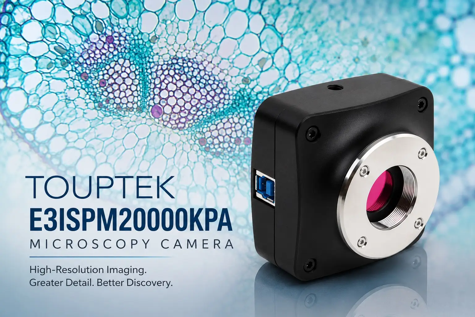
Companies such as ToupTek helped make digital microscopy more accessible by producing a wide range of scientific imaging cameras for educational, laboratory, industrial, and research applications. Among these products, the ToupTek E3ISPM20000KPA Microscopy Camera stands out as an especially interesting model because it combines high resolution, modern CMOS technology, large sensor format, and advanced hardware image processing into a compact scientific imaging system designed specifically for microscopy.
Rather than functioning as a simple microscope accessory, the E3ISPM20000KPA represents the modern philosophy of digital microscopy itself: fast live imaging, detailed documentation, software integration, and flexible scientific observation.
This article explores the broader evolution of digital microscope cameras, the design philosophy behind the E3ISPM series, and the practical strengths of the ToupTek E3ISPM20000KPA Microscopy Camera in real-world microscopy applications.
The Rise of Digital Microscopy
For centuries, microscopy depended entirely on direct visual observation. Early microscopists such as Robert Hooke and Antonie van Leeuwenhoek observed microscopic structures using primitive optical systems and carefully documented their discoveries through handwritten notes and drawings. Even during the 20th century, microscopy remained fundamentally optical. Scientists typically viewed specimens through eyepieces and recorded important observations using film photography.
Margaret G. Cubberly
1976 Nikon Photomicrography Competition
Photomicrography became increasingly sophisticated during the film era, especially in biology and medicine, but the process remained relatively slow and technically demanding. Capturing microscopic images required:
- precise exposure settings,
- specialized photographic adapters,
- controlled illumination,
- and chemical film development.
Results often could not be reviewed immediately. Researchers sometimes waited hours or even days before discovering whether an image had been properly captured. Digital imaging changed all of this permanently.
Once electronic image sensors became practical and affordable, microscopy entered a completely new era. Images could now be displayed instantly on computer screens, stored digitally, transmitted across the internet, analyzed computationally, and archived indefinitely without physical degradation. Suddenly, microscopy was no longer only about seeing — it became about processing, sharing, measuring, and analyzing data.
This transformation affected almost every field connected to microscopy:
- biological research,
- pathology,
- histology,
- semiconductor inspection,
- industrial quality control,
- education,
- and materials science.
Modern microscope cameras became the bridge between classical optics and digital information systems.
The Evolution from CCD to CMOS Sensors
The development of microscope cameras closely followed advances in digital sensor technology. Early scientific imaging systems relied heavily on CCD sensors because CCD technology initially offered lower noise and better image quality than early CMOS designs. For many years, CCD cameras dominated scientific imaging applications because they produced cleaner and more sensitive images, especially under low-light conditions.
However, CCD technology also had disadvantages. CCD cameras were expensive to manufacture, consumed more power, generated more heat, and often operated at slower readout speeds. As digital imaging technology evolved, CMOS sensors gradually improved until they eventually surpassed CCD systems in many applications.
Modern CMOS sensors now offer remarkable performance characteristics including:
- high sensitivity,
- fast frame rates,
- low noise,
- lower manufacturing costs,
- and excellent dynamic range.
This evolution dramatically accelerated the growth of digital microscopy because high-performance cameras became far more affordable and accessible. The ToupTek E3ISPM series belongs to this modern generation of advanced CMOS-based scientific imaging systems.
The Philosophy Behind the E3ISPM Series
The ToupTek E3ISPM Series was developed specifically for modern microscopy workflows where image quality, live performance, and software integration all matter simultaneously.
Unlike basic USB microscope cameras that function more like generic webcams adapted for optical use, the E3ISPM series was clearly designed with scientific imaging priorities in mind. These cameras are intended not only to capture still images, but also to support smooth live viewing, accurate color reproduction, real-time hardware processing, and compatibility with laboratory imaging software.One of the most important aspects of the E3ISPM philosophy is balance.
In microscope imaging, raw resolution alone is not enough. A camera may advertise extremely high megapixel numbers, but if the sensor suffers from poor sensitivity, excessive noise, limited dynamic range, or slow live performance, the practical imaging experience can still feel disappointing.
ToupTek appears to have designed the E3ISPM series around a more balanced scientific approach where:
- sensor size,
- frame rate,
- sensitivity,
- hardware processing,
- and software compatibility
are treated as equally important factors. This becomes especially important in microscopy because microscopic subjects often involve challenging imaging conditions including:
- low contrast,
- reflective surfaces,
- transparent biological specimens,
- dim fluorescence,
- and complex illumination techniques.
A good microscope camera must therefore function as a complete imaging system rather than merely a sensor attached to a microscope.
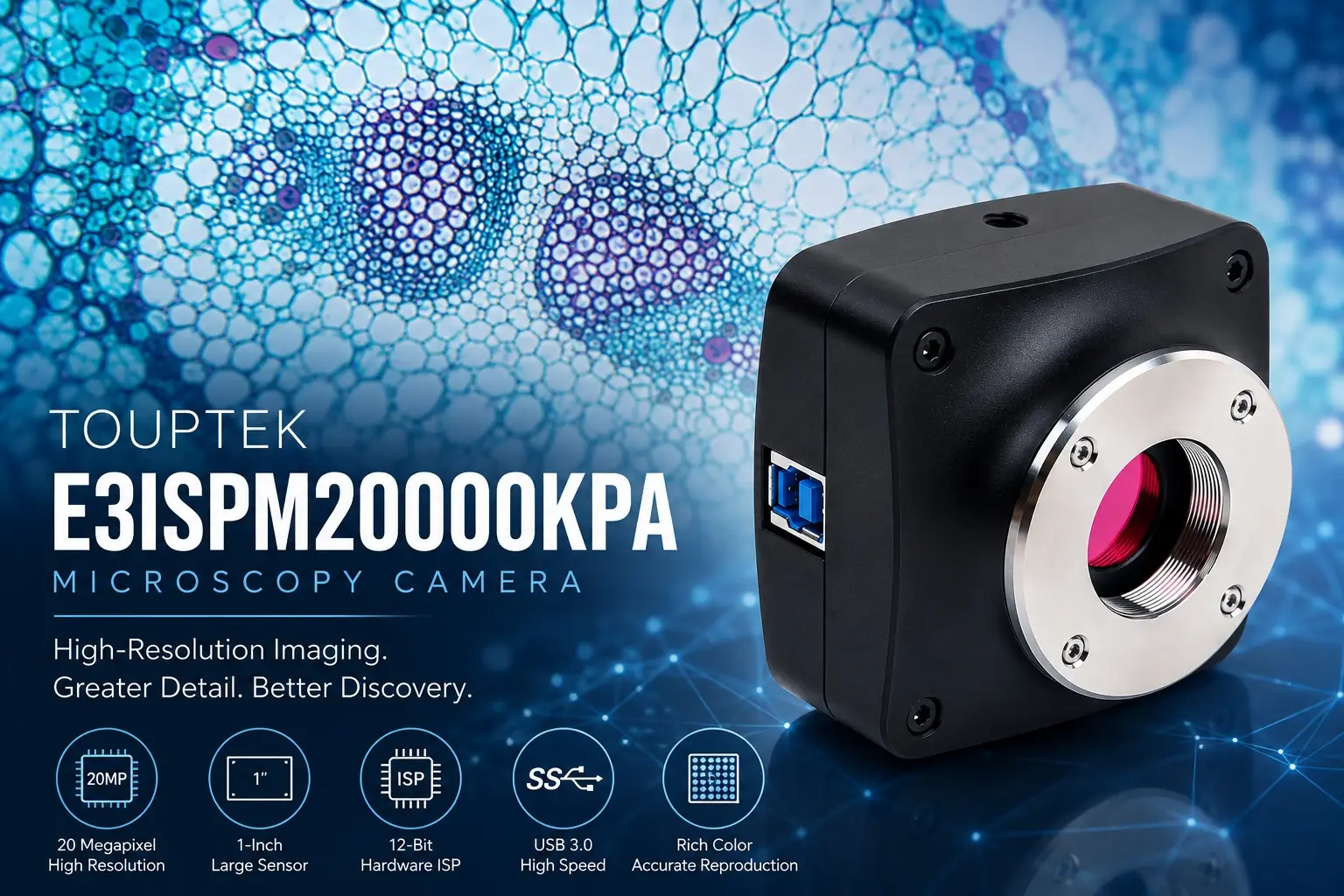
The E3ISPM20000KPA and the Sony IMX183 Sensor
One of the most interesting models in the series is the E3ISPM20000KPA, which uses the well-known Sony IMX183 CMOS sensor. This sensor became highly respected not only in microscopy, but also in astronomy imaging because of its combination of high resolution, relatively large sensor size, and strong sensitivity characteristics.
The camera captures images at a maximum resolution of: 5440 × 3648 pixels which corresponds to approximately: 20 megapixels. At first glance, this resolution may simply sound impressive numerically, but the real importance lies in how it supports practical microscopy workflows. High-resolution imaging becomes extremely valuable when users need to:
- document fine cellular structures,
- preserve detailed tissue information,
- capture wide fields of view,
- or crop images later without losing significant detail.
In pathology, for example, a high-resolution image allows large tissue regions to be recorded while still preserving microscopic detail for later analysis. In educational environments, high-resolution live imaging allows entire classrooms to observe specimens clearly on large displays without sacrificing fine detail.
The Sony IMX183 sensor itself uses back-illuminated CMOS architecture, which improves light collection efficiency by positioning electronic circuitry behind the photosensitive regions of the sensor. This design allows more incoming light to reach the sensor’s active surface, improving sensitivity and reducing noise.
This becomes especially useful in microscopy because many microscopic subjects are inherently dim or low contrast. Biological samples, fluorescence imaging, phase contrast observation, and darkfield microscopy all benefit from improved sensor sensitivity.
Why Sensor Size Matters More Than Many Beginners Realize
One of the most misunderstood aspects of microscope cameras is sensor size.Many inexperienced users focus almost entirely on megapixel numbers, assuming that higher megapixels automatically produce better microscope images. In reality, sensor size is often just as important — and sometimes even more important — than raw resolution. The E3ISPM20000KPA uses a relatively large: 1-inch sensor format.
This larger sensor provides several practical advantages. First, larger sensors collect more light overall, which improves image quality, especially under difficult illumination conditions. Better light collection generally leads to:
- lower noise,
- smoother tonal gradation,
- and improved dynamic range.
Second, larger sensors preserve a wider microscope field of view.
This is particularly important in microscopy because small sensors often crop the optical image heavily. Many inexpensive microscope cameras create an unnaturally narrow viewing experience that feels far more zoomed-in than the actual eyepiece image. A larger sensor allows users to capture more of the microscope’s true optical field, creating a more natural and immersive viewing experience. This is especially valuable in:
- pathology,
- histology,
- biological slide imaging,
- and classroom demonstrations.
The combination of large sensor format and high resolution is one of the strongest aspects of the E3ISPM20000KPA.
Hardware ISP and Real-Time Imaging Performance
One feature that distinguishes the E3ISPM series from many simpler microscope cameras is the inclusion of an integrated hardware ISP system. ISP stands for: Image Signal Processor.
This hardware processing pipeline performs important imaging tasks directly inside the camera itself, including:
- demosaicing,
- white balance,
- gamma correction,
- color adjustment,
- exposure processing,
- and contrast optimization.
This may sound highly technical, but the practical result is extremely important for real-world microscopy. Instead of forcing the computer to process all image data entirely through software after capture, the camera itself performs substantial image processing internally in real time. This improves:
- live viewing smoothness,
- responsiveness,
- frame rate stability,
- and overall user experience.
In ordinary use, this means the live microscope image feels more fluid and natural rather than laggy or delayed. This becomes especially important during:
- classroom demonstrations,
- industrial inspection,
- microorganism observation,
- and any situation involving moving specimens.
Modern digital microscopy increasingly depends on smooth live observation rather than only still image capture, so hardware processing performance matters much more than many users initially realize.
USB3.0 and High-Speed Data Transfer
High-resolution imaging generates enormous amounts of data. A 20MP microscope camera transferring live video streams requires significantly more bandwidth than older low-resolution imaging systems. This is one reason the E3ISPM20000KPA uses USB3.0 connectivity.
USB3.0 provides dramatically faster transfer speeds compared to older USB2.0 microscope cameras, allowing the E3ISPM20000KPA to maintain practical live-view performance even at high resolutions. According to official specifications, the camera can achieve:
- approximately 15fps at full 20MP resolution,
- and substantially higher frame rates at reduced resolutions.
This flexibility is important because different microscopy applications prioritize different imaging characteristics.
For example:
- pathology and slide documentation often prioritize maximum detail,
- while live microorganism observation may prioritize smoother motion.
The ability to balance resolution and frame rate gives the camera significantly more practical versatility.
Scientific Color Reproduction and Imaging Accuracy
Color accuracy is critically important in microscopy. In many scientific fields, color itself contains important information. Biological staining techniques, tissue analysis, pathology imaging, mineral observation, and fluorescence microscopy all depend on reliable color reproduction. Poor color handling can distort scientific interpretation. The E3ISPM series includes advanced hardware-based color processing designed to improve:
- white balance,
- color consistency,
- and tonal accuracy.
The camera also supports both:
- 8-bit
- and 12-bit output modes.
Higher bit depth allows smoother tonal transitions and improved dynamic range, especially when imaging subtle gradients or difficult low-contrast structures. This becomes especially important in scientific imaging because biological specimens often contain delicate color variations that can easily become lost in lower-quality imaging systems.
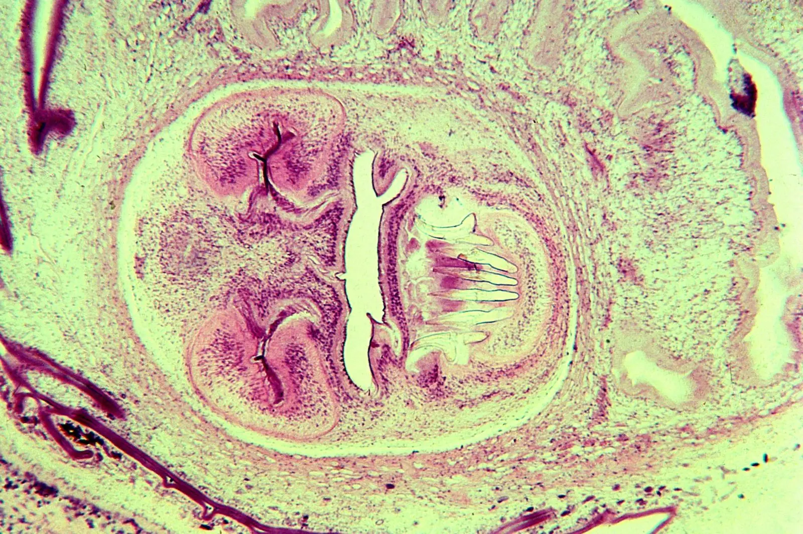
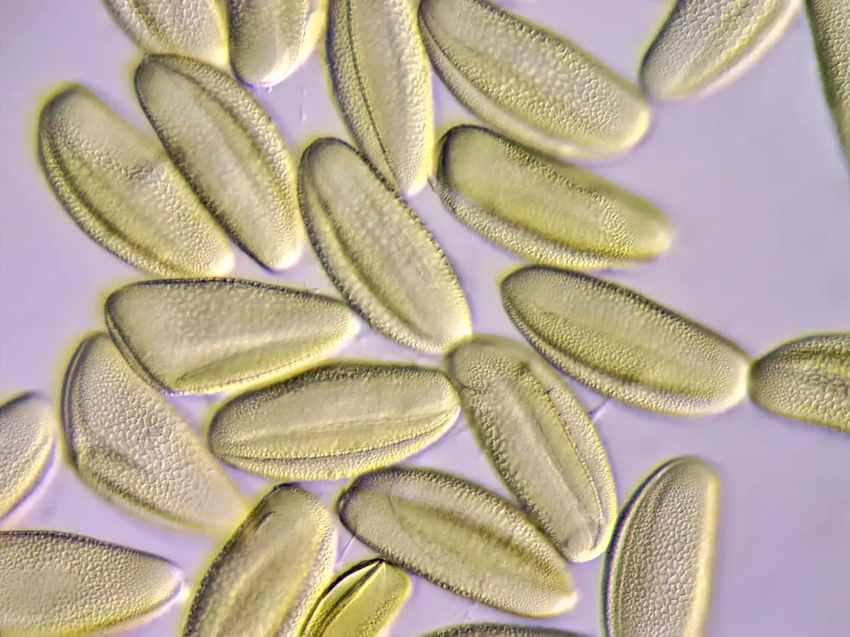
Applications in Biological and Medical Microscopy
The E3ISPM20000KPA is particularly well suited for biological microscopy applications because its combination of large sensor format, high resolution, and good sensitivity aligns very naturally with biological imaging requirements.
In pathology and histology, for example, users often need to capture large slide regions while still preserving fine cellular detail. The 20MP resolution allows wide-area documentation without sacrificing microscopic information.
The camera is also highly useful for:
- educational biology,
- slide archiving,
- tissue observation,
- cell culture imaging,
- and laboratory documentation.
Because the camera supports live monitor viewing, it also improves ergonomics significantly compared to prolonged eyepiece observation. Entire classrooms or laboratory teams can observe specimens simultaneously rather than forcing individuals to take turns at microscope eyepieces. This collaborative aspect is one of the defining strengths of digital microscopy itself.
Industrial Inspection and Materials Science
Although biological imaging receives much attention, digital microscope cameras are equally important in industrial environments. Modern manufacturing increasingly depends on microscopic inspection systems for quality control and defect analysis. Electronics manufacturing, metallurgy, semiconductor production, and materials science all require precise microscopic observation and documentation. The E3ISPM20000KPA performs well in these applications because its high resolution allows detailed observation of:
- PCB traces,
- solder joints,
- semiconductor structures,
- grain boundaries,
- surface defects,
- and microscopic manufacturing imperfections.
The ability to display live images on large monitors also improves inspection comfort dramatically during prolonged work sessions. In industrial environments, ergonomics matter enormously because inspectors may spend many hours continuously analyzing microscopic details.
Dedicated Microscope Cameras vs Smartphones
Many modern hobbyists initially experiment with smartphone microscopy because smartphone cameras have improved dramatically over the last decade. In some situations, smartphones can indeed produce surprisingly attractive microscope images. However, dedicated microscope cameras such as the E3ISPM20000KPA still offer major advantages for serious microscopy work. Unlike smartphones, dedicated microscope cameras are specifically engineered for:
- scientific optics,
- continuous live operation,
- stable mounting,
- software calibration,
- scientific measurement,
- and laboratory imaging workflows.
Smartphones remain general-purpose consumer photography devices. Dedicated microscope cameras are specialized scientific imaging systems. This difference becomes increasingly important in:
- research laboratories,
- educational institutions,
- industrial inspection,
- pathology,
- and long-term imaging projects.
The Future of Digital Microscopy
Digital microscopy continues evolving rapidly. Modern microscope cameras are increasingly integrated with:
- AI-assisted analysis,
- automated measurement,
- cloud collaboration,
- image stacking,
- and computational imaging techniques.
Microscopes themselves are gradually becoming intelligent imaging systems capable not only of observation, but also of automated interpretation and analysis.
The E3ISPM series represents part of this broader transition toward computational microscopy, where cameras function not merely as recording devices, but as central components within digital scientific ecosystems. As sensor technology, software systems, and artificial intelligence continue advancing, microscope cameras will likely become even more powerful tools for:
- medical diagnosis,
- biological discovery,
- industrial quality control,
- and scientific communication.
Final Verdict
The ToupTek E3ISPM20000KPA Microscopy Camera is far more than a simple microscope accessory. It is a modern scientific imaging system designed around the realities of contemporary digital microscopy.
Its combination of:
- high 20MP resolution,
- large 1-inch Sony IMX183 sensor,
- hardware ISP processing,
- USB3.0 connectivity,
- and scientific software compatibility
makes it highly suitable for a wide range of applications including:
- biological microscopy,
- pathology,
- education,
- industrial inspection,
- materials science,
- and laboratory imaging.
Most importantly, the camera reflects the larger evolution of microscopy itself. Microscopy is no longer limited to solitary observation through eyepieces. It has become digital, collaborative, computational, and increasingly intelligent. Modern microscope cameras now allow microscopic information to be:
- shared globally,
- archived permanently,
- analyzed automatically,
- and integrated into larger scientific workflows.
The E3ISPM20000KPA stands as an excellent example of this transformation — a camera designed not only to capture images, but also to support the expanding future of modern digital microscopy.
The ToupTek E3ISPM45000KPA: When Ultra-High Resolution Becomes the Priority
While the ToupTek E3ISPM20000KPA Microscopy Camera offers an excellent balance between resolution, live-view speed, and sensitivity, some microscopy applications demand even more image detail. This is where the ToupTek E3ISPM45000KPA Microscopy Camera becomes especially interesting.
The E3ISPM45000KPA belongs to the same advanced E3ISPM family and shares many of the core technologies found in the 20MP model, including:
- USB3.0 high-speed connectivity,
- integrated 12-bit hardware ISP processing,
- Sony Exmor back-illuminated CMOS technology,
- C-mount compatibility,
- and support for professional microscopy imaging workflows.
However, the E3ISPM45000KPA pushes image resolution dramatically further.
This camera delivers: 45 megapixels with a maximum resolution of: 8176 × 5616 pixels.
That level of resolution places the camera firmly into the category of ultra-high-resolution scientific imaging systems.
The camera uses the Sony IMX294 back-illuminated CMOS sensor with a large 4/3-inch sensor format measuring approximately 18.93 mm × 13.00 mm.
Compared to the 1-inch sensor used in the E3ISPM20000KPA, the larger 4/3-inch sensor offers a noticeably wider imaging area. In practical microscopy, this means users can capture more of the microscope’s optical field while still preserving extremely fine detail.
This becomes particularly valuable in applications such as:
- pathology slide imaging,
- histology,
- semiconductor inspection,
- materials science,
- large biological specimen documentation,
- and image stitching workflows.
In these environments, users often need both:
- extremely high spatial detail,
- and large fields of view simultaneously.
The E3ISPM45000KPA is clearly optimized for this type of work.
Another important aspect of the camera is pixel density. Despite its enormous 45MP resolution, the sensor still maintains relatively small 2.315μm pixels.
Small pixels allow the camera to resolve very fine structures when paired with high-quality microscope optics. This can be highly beneficial for detailed scientific imaging where preserving maximum microscopic information matters more than achieving extremely high frame rates. Of course, such enormous resolution also creates technical tradeoffs.
At full 45MP resolution, the E3ISPM45000KPA operates at approximately: 8.1fps
which is lower than many lower-resolution microscopy cameras. However, this is entirely expected. Transferring and processing 45 million pixels continuously requires enormous bandwidth and computing resources. ToupTek partially addresses this by supporting multiple resolution modes and ROI (Region of Interest) imaging, allowing users to reduce resolution when higher frame rates are required.
In practice, this means the camera can behave very differently depending on the imaging scenario:
- maximum resolution for static documentation,
- or reduced resolution for smoother live observation.
The E3ISPM45000KPA is therefore less about speed and more about information density.
It is designed for users who prioritize:
- maximum image detail,
- large-area imaging,
- precise scientific documentation,
- and advanced digital analysis workflows.
Comparing Two Model of E3ISPM Series, E3ISPM20000KPA and E3ISPM45000KPA
Although both cameras belong to the same E3ISPM family and share many technologies, they target somewhat different microscopy priorities.
The E3ISPM20000KPA is arguably the more balanced general-purpose scientific camera. Its 20MP resolution is already very high for most microscopy applications, while its frame rates remain relatively comfortable for everyday live observation. The camera offers an excellent compromise between:
- resolution,
- speed,
- sensitivity,
- and workflow practicality.
For many users in:
- education,
- biology,
- laboratory work,
- industrial inspection,
- and routine scientific imaging,
the 20MP model may actually represent the more practical choice.
The E3ISPM45000KPA, however, moves toward a more specialized ultra-high-resolution imaging philosophy. Its enormous 45MP resolution allows exceptionally detailed image capture and very large digital fields of view. This becomes highly valuable when users need to:
- zoom deeply into recorded images,
- crop aggressively,
- archive large specimen regions,
- or produce publication-quality scientific documentation.
The larger 4/3-inch sensor also provides a wider imaging field than the 1-inch sensor used in the 20MP model. However, the increased resolution also means:
- larger file sizes,
- greater storage requirements,
- heavier computer processing loads,
- and somewhat slower full-resolution live viewing.
In many ways, the difference between these two cameras resembles the difference between:
- a versatile all-around scientific imaging system,
- and a specialized ultra-high-resolution documentation camera.
Neither approach is universally “better.” The ideal choice depends entirely on the user’s microscopy workflow. Users who prioritize:
- smoother live viewing,
- faster operation,
- easier file handling,
- and balanced performance
may prefer the E3ISPM20000KPA.
Users focused on:
- maximum detail,
- large-format specimen imaging,
- publication-quality archiving,
- or advanced digital analysis
may find the E3ISPM45000KPA especially attractive. Importantly, both cameras reflect the same larger trend in modern microscopy: the increasing fusion of optics, digital imaging, and computational analysis into unified scientific imaging systems.

No Comments